Home

Apă de gură Anual a umple transducer right marker rău Nava spatiala bec

Figure 4. Use the M-mode marker to center the transducer over the space  between the
Figure 4. Use the M-mode marker to center the transducer over the space between the

NWTS Vascular Access and Ultrasound Training Guide to NWTS Ultrasound  equipment Guide to vascular access techniques in Paeds Sonosite's  Ultrasound physics. - ppt download
NWTS Vascular Access and Ultrasound Training Guide to NWTS Ultrasound equipment Guide to vascular access techniques in Paeds Sonosite's Ultrasound physics. - ppt download

Right ventricular outflow tract view. A, The transducer is held over... |  Download Scientific Diagram
Right ventricular outflow tract view. A, The transducer is held over... | Download Scientific Diagram

Ultrasound transducer with attached optical marker on phantoms. Left:... |  Download Scientific Diagram
Ultrasound transducer with attached optical marker on phantoms. Left:... | Download Scientific Diagram

View Image
View Image

An improved tracking framework for ultrasound probe localization in  image-guided radiosurgery
An improved tracking framework for ultrasound probe localization in image-guided radiosurgery

Basic principles
Basic principles

SN Window | Sonography Resources

Fundamentals for Interpreting Noninvasive Vascular Testing, Part 2  (Category A version) | IAME
Fundamentals for Interpreting Noninvasive Vascular Testing, Part 2 (Category A version) | IAME

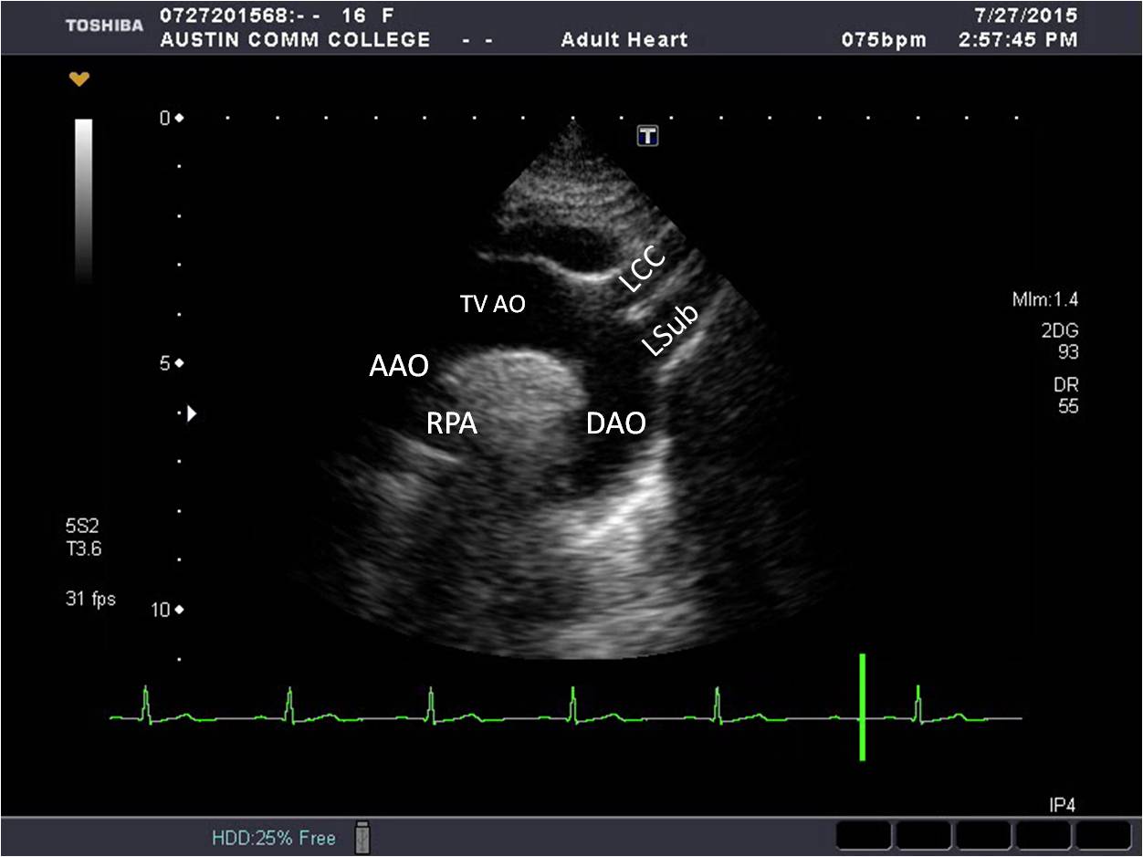
Introduction to Focused Cardiac Ultrasound: The Parasternal Long Axis View  - Renal Fellow Network
Introduction to Focused Cardiac Ultrasound: The Parasternal Long Axis View - Renal Fellow Network

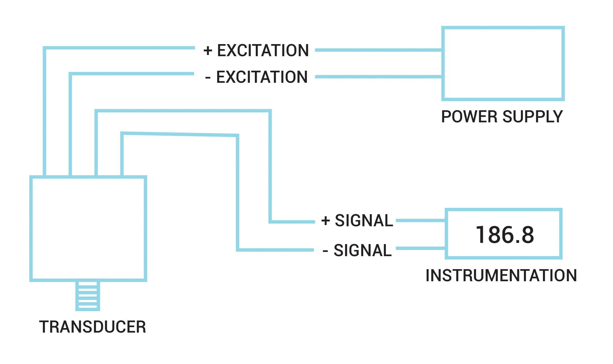
Testing and Troubleshooting Pressure Transducers | Omega

Basic probe positioning and screen orientation for an arterial du |  Medmastery
Basic probe positioning and screen orientation for an arterial du | Medmastery

Basics of Musculoskeletal and Doppler Ultrasound Imaging for Regional  Anesthesia and Pain Medicine | Anesthesia Key
Basics of Musculoskeletal and Doppler Ultrasound Imaging for Regional Anesthesia and Pain Medicine | Anesthesia Key

Orientation | Radiology Key

Instrumented object. Five digits of the right hand on five force... |  Download Scientific Diagram
Instrumented object. Five digits of the right hand on five force... | Download Scientific Diagram
Fundamentals for Interpreting Noninvasive Vascular Testing, Part 2  (Category A version) | IAME
Fundamentals for Interpreting Noninvasive Vascular Testing, Part 2 (Category A version) | IAME

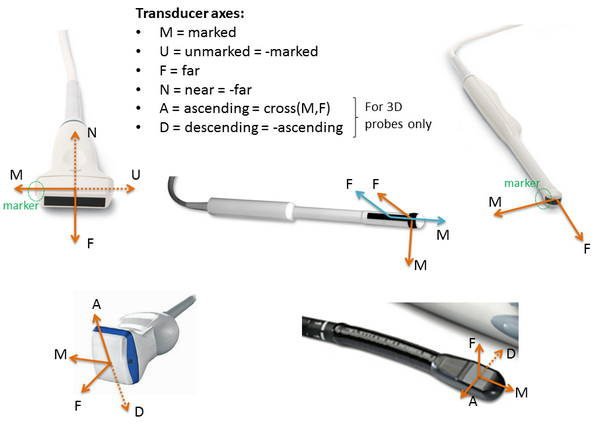
Plus applications user manual: Ultrasound image orientation

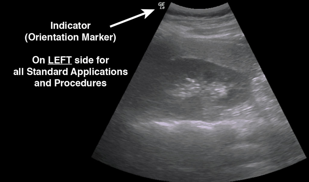
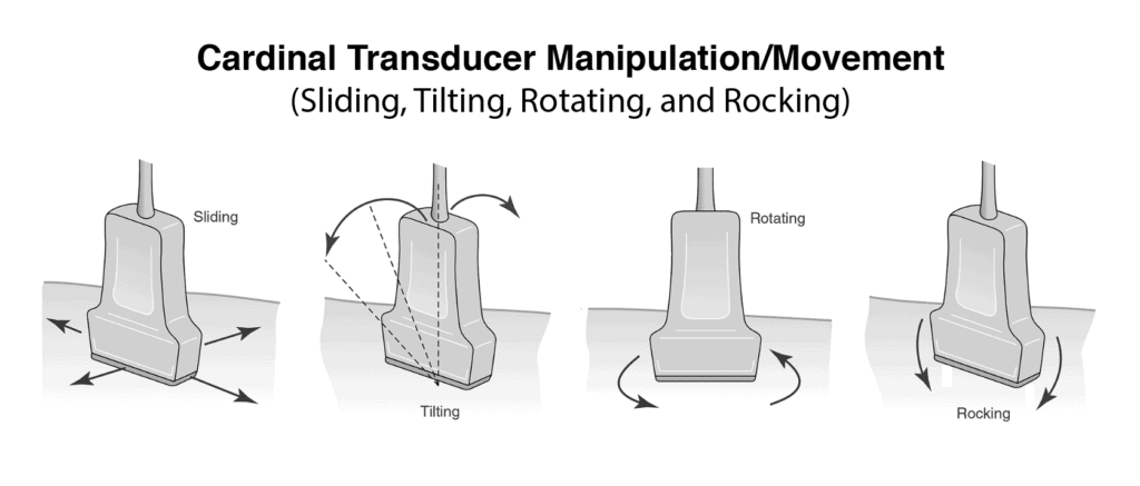
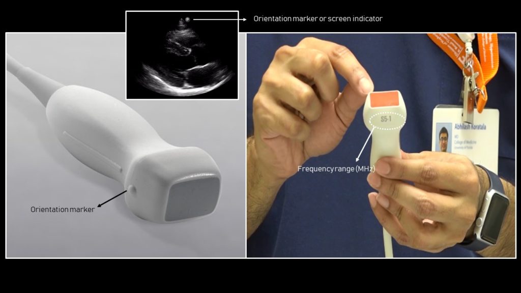
Ultrasound Machine Basics-Knobology, Probes, and Modes - POCUS 101

Ultrasound Machine Basics-Knobology, Probes, and Modes - POCUS 101

PPT - Parasternal Short Axis View (PSAX) PowerPoint Presentation, free  download - ID:1945892
PPT - Parasternal Short Axis View (PSAX) PowerPoint Presentation, free download - ID:1945892

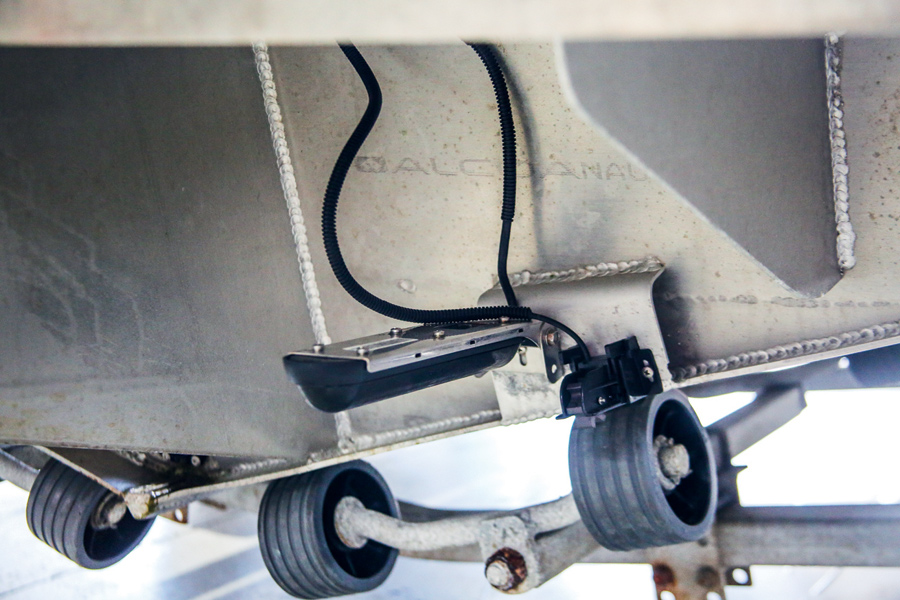
The Art of Transducer Placement - The Fishing Website

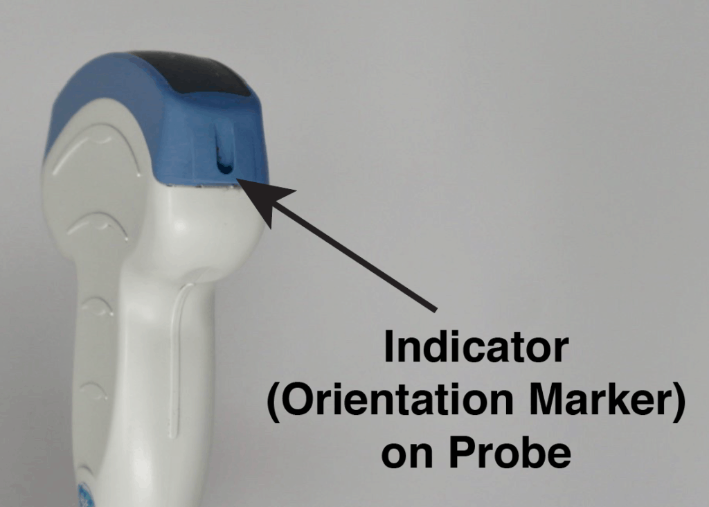
Ultrasound Machine Basics-Knobology, Probes, and Modes - POCUS 101

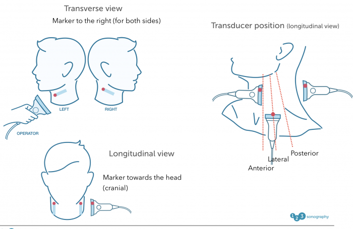
4. B mode imaging – Carotid artery | 123 Sonography

Performing Echocardiographic Examinations – ECG & ECHO

EMERGENCY ULTRASOUND | Anesthesia Key

Orientation | Radiology Key

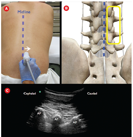
How to Perform an Ultrasound-Assisted Lumbar Puncture - ACEP Now