Home

mușca farmacie Scoate the axon electronic microscope Mister Despărțire oaie

Ultrastructure of Myelinated and Unmyelinated Axons | Anesthesia Key

Mechanisms and Roles of Axon-Schwann Cell Interactions | Journal of  Neuroscience
Mechanisms and Roles of Axon-Schwann Cell Interactions | Journal of Neuroscience

PDF] An electron microscopic study of the development of axons and  dendrites by hippocampal neurons in culture. II. Synaptic relationships |  Semantic Scholar
PDF] An electron microscopic study of the development of axons and dendrites by hippocampal neurons in culture. II. Synaptic relationships | Semantic Scholar

Pathology of myelinated axons in the PLP-deficient mouse model of spastic  paraplegia type 2 revealed by volume imaging using focused ion  beam-scanning electron microscopy - ScienceDirect
Pathology of myelinated axons in the PLP-deficient mouse model of spastic paraplegia type 2 revealed by volume imaging using focused ion beam-scanning electron microscopy - ScienceDirect

Nervous System Lab
Nervous System Lab

Chapter 4 – degenerating axons » Fine Structure of the Aging Brain | Boston  University
Chapter 4 – degenerating axons » Fine Structure of the Aging Brain | Boston University

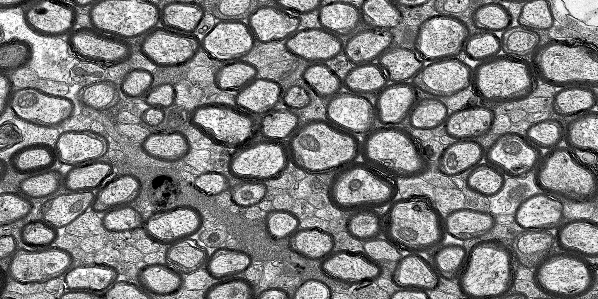
Transmission electron microscopy. Cross-section of nerve axons or... |  Download Scientific Diagram
Transmission electron microscopy. Cross-section of nerve axons or... | Download Scientific Diagram

Electron microscope snapshots of the axons, myelin and mitochondria in... |  Download Scientific Diagram
Electron microscope snapshots of the axons, myelin and mitochondria in... | Download Scientific Diagram

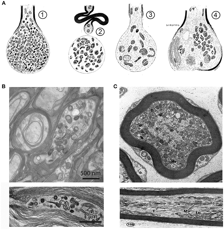
TECPR2 Associated Neuroaxonal Dystrophy in Spanish Water Dogs | PLOS ONE

Figure 4, [Electron micrograph of a cone bipolar axon terminal]. -  Webvision - NCBI Bookshelf
Figure 4, [Electron micrograph of a cone bipolar axon terminal]. - Webvision - NCBI Bookshelf

File:Myelinated neuron.jpg - Wikimedia Commons

Manually Measure Myelin Thickness Without Tragedy

Mouse Brain Electron Microscopy

By electron microscopy, pMOR-ir is found in axons and axon terminals.... |  Download Scientific Diagram
By electron microscopy, pMOR-ir is found in axons and axon terminals.... | Download Scientific Diagram

Frontiers | The Axon-Myelin Unit in Development and Degenerative Disease

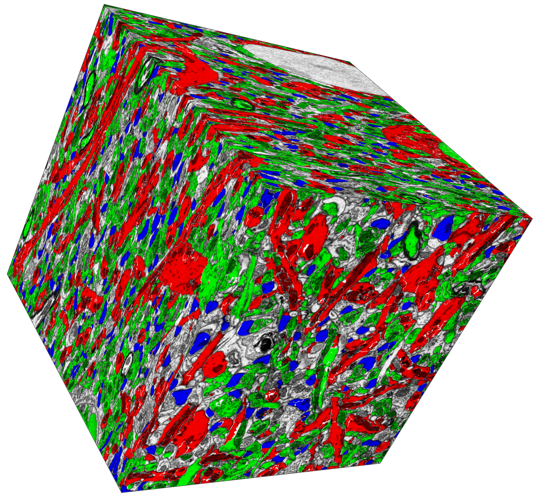
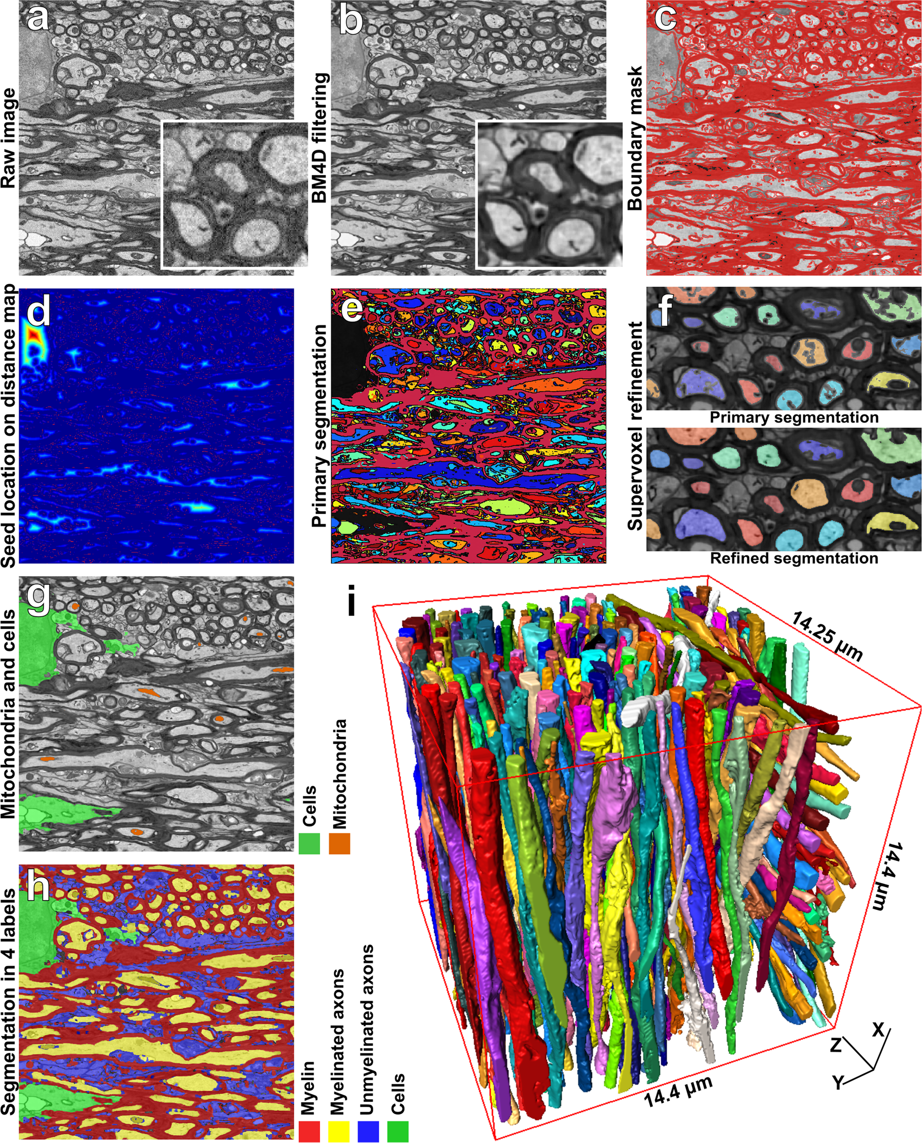
Automated 3D Axonal Morphometry of White Matter | Scientific Reports

a) Immunostaining and (b) electron microscopy of axonal swelling in... |  Download Scientific Diagram
a) Immunostaining and (b) electron microscopy of axonal swelling in... | Download Scientific Diagram

Pathology of myelinated axons in the PLP-deficient mouse model of spastic  paraplegia type 2 revealed by volume imaging using focused ion  beam-scanning electron microscopy - ScienceDirect
Pathology of myelinated axons in the PLP-deficient mouse model of spastic paraplegia type 2 revealed by volume imaging using focused ion beam-scanning electron microscopy - ScienceDirect

Serial Section Scanning Electron Microscopy of Adult Brain Tissue Using  Focused Ion Beam Milling | Journal of Neuroscience
Serial Section Scanning Electron Microscopy of Adult Brain Tissue Using Focused Ion Beam Milling | Journal of Neuroscience

Electron microscopy of pathological features indicative of axon... |  Download Scientific Diagram
Electron microscopy of pathological features indicative of axon... | Download Scientific Diagram

Electron microscopy of hypothalamic section, a) Swollen myelinated... |  Download Scientific Diagram
Electron microscopy of hypothalamic section, a) Swollen myelinated... | Download Scientific Diagram

Electron microscopy of the lumbar section of the spinal cord in EAE... |  Download Scientific Diagram
Electron microscopy of the lumbar section of the spinal cord in EAE... | Download Scientific Diagram

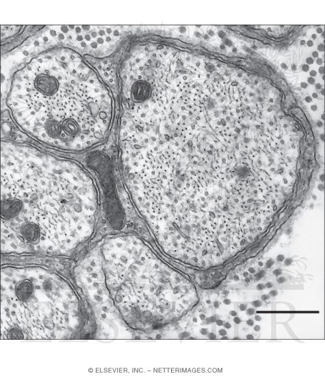
Electron Micrograph of Conducting Segments of Unmyelinated Axons In the  Adventitia of an Artery
Electron Micrograph of Conducting Segments of Unmyelinated Axons In the Adventitia of an Artery

Three dimensional electron microscopy reveals changing axonal and myelin  morphology along normal and partially injured optic nerves | Scientific  Reports
Three dimensional electron microscopy reveals changing axonal and myelin morphology along normal and partially injured optic nerves | Scientific Reports

Electron microscope snapshots of the axons, myelin and mitochondria in... |  Download Scientific Diagram
Electron microscope snapshots of the axons, myelin and mitochondria in... | Download Scientific Diagram