Home

ziarist supă Pronume skull mri bone anatomy lamina spalare listă Profetia

Geometric and mechanical evaluation of 3D-printing materials for skull base  anatomical education and endoscopic surgery simulation – A first step to  create reliable customized simulators | PLOS ONE
Geometric and mechanical evaluation of 3D-printing materials for skull base anatomical education and endoscopic surgery simulation – A first step to create reliable customized simulators | PLOS ONE

Imaging of the Anterior Skull Base | Radiology Key

Imaging review of the anterior skull base - Olivia Francies, Levan  Makalanda, Dimitris Paraskevopolous, Ashok Adams, 2018
Imaging review of the anterior skull base - Olivia Francies, Levan Makalanda, Dimitris Paraskevopolous, Ashok Adams, 2018

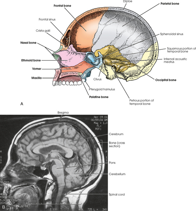
Cranium and Facial Bones - Sectional anatomy for imaging professionals, 4th  edition
Cranium and Facial Bones - Sectional anatomy for imaging professionals, 4th edition

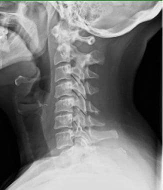
Cervical Spine Anatomy: Overview, Gross Anatomy

Skull: Anatomy, structure, bones, quizzes | Kenhub

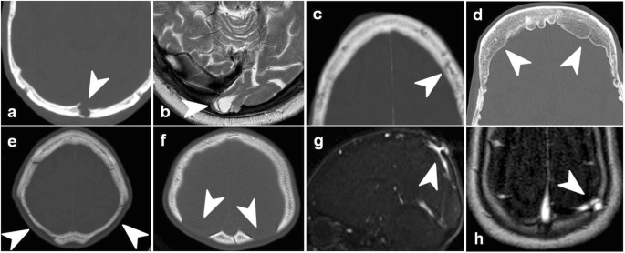
Imaging of skull vault tumors in adults | Insights into Imaging | Full Text

A) Sagittal, (B) coronal, and (C) axial T1 weighted magnetic resonance... |  Download Scientific Diagram
A) Sagittal, (B) coronal, and (C) axial T1 weighted magnetic resonance... | Download Scientific Diagram
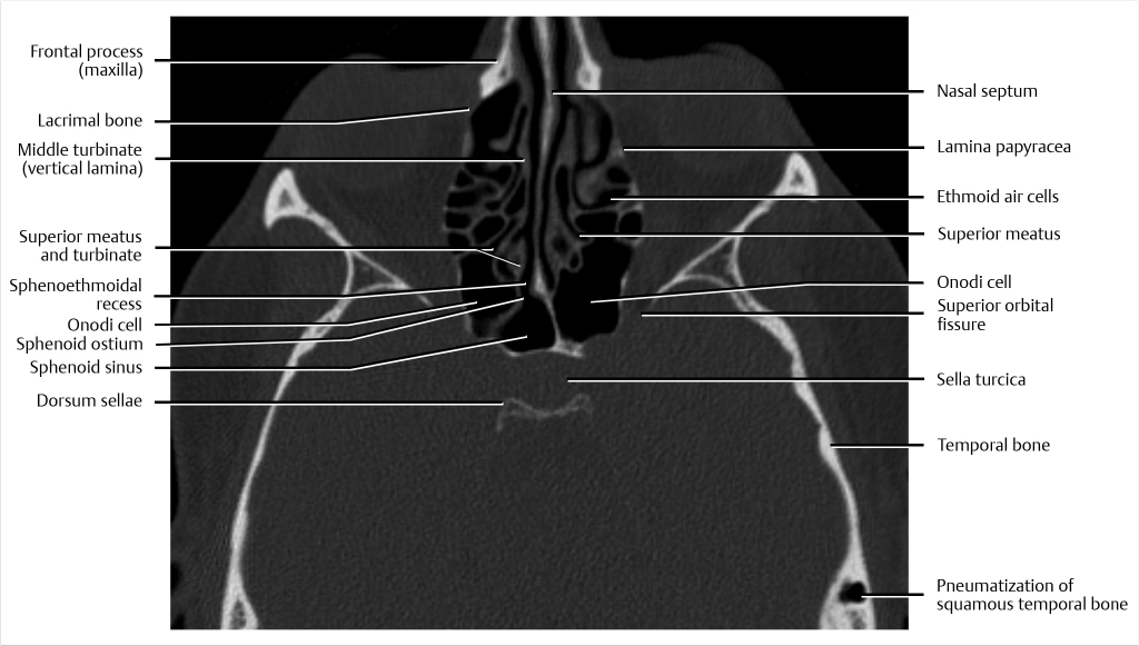
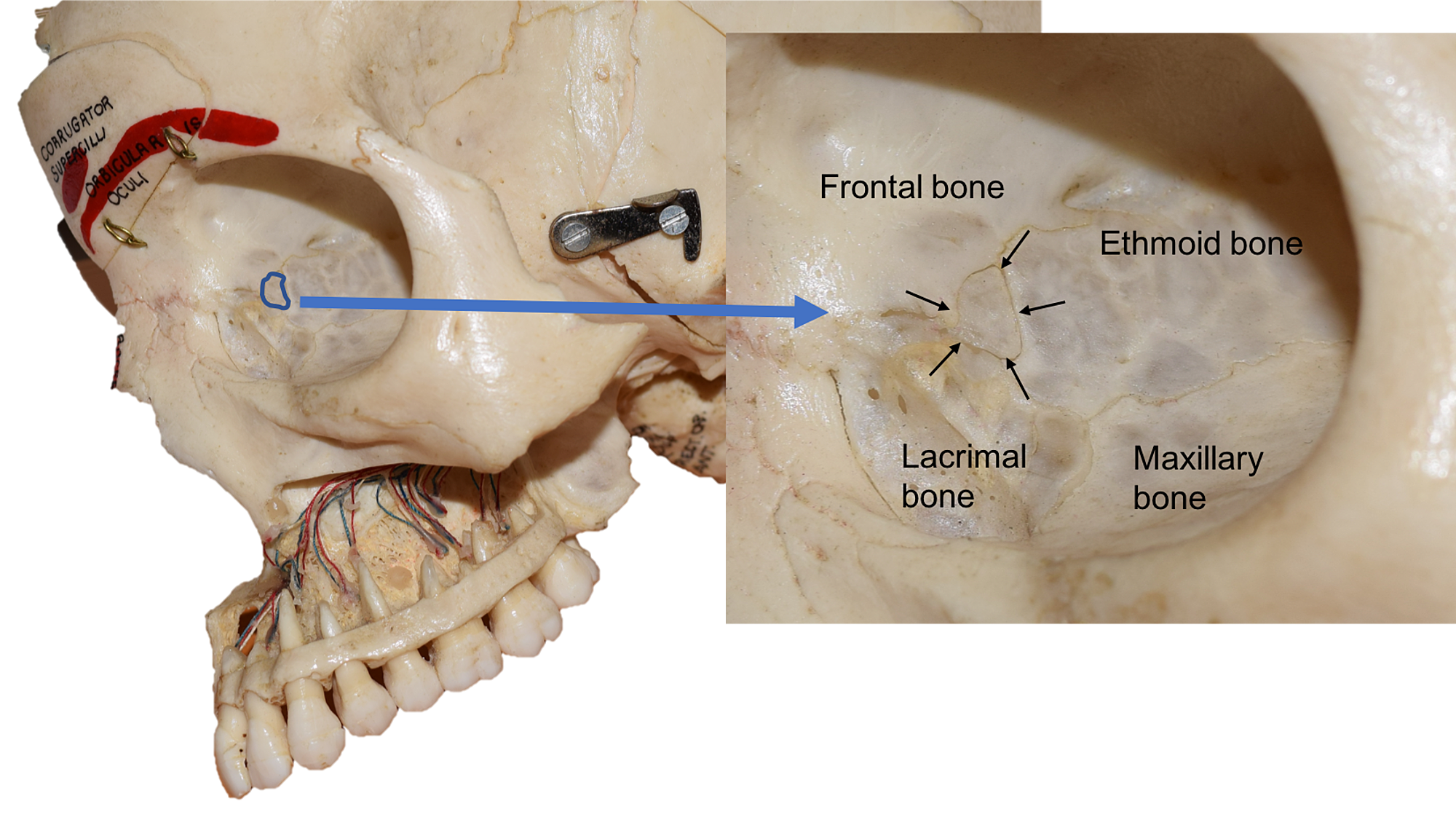
Cureus | Wormian Bone of the Orbit: A Case Report

Cross-Sectional Computed Tomography and Magnetic Resonance Imaging Atlas of  the Skull Base | Radiology Key
Cross-Sectional Computed Tomography and Magnetic Resonance Imaging Atlas of the Skull Base | Radiology Key

Normal anatomy of the base of the skull, orbit, pituitary and cranial  nerves | Practical Neurology
Normal anatomy of the base of the skull, orbit, pituitary and cranial nerves | Practical Neurology

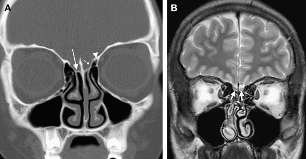
Case 1. (A) Coronal T1 magnetic resonance imaging (MRI) and (B) coronal...  | Download Scientific Diagram
Case 1. (A) Coronal T1 magnetic resonance imaging (MRI) and (B) coronal... | Download Scientific Diagram

MIDA: A Multimodal Imaging-Based Detailed Anatomical Model of the Human Head  and Neck | PLOS ONE
MIDA: A Multimodal Imaging-Based Detailed Anatomical Model of the Human Head and Neck | PLOS ONE

A comprehensive review of the imaging of skull base infections – Part I -  ScienceDirect
A comprehensive review of the imaging of skull base infections – Part I - ScienceDirect

Superior view of the base of the skull: Anatomy | Kenhub

Lumbar spine anatomy on MRI (Magnetic Resonance Imaging) - e-Anatomy

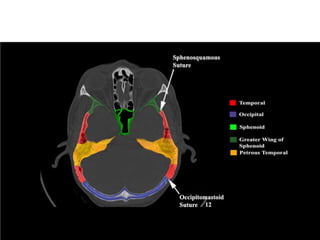
Imaging of skull base: Pictorial essay. - Abstract - Europe PMC

Topography of The Skull: Internal surface of the cranial base

Topography of The Skull: Internal surface of the cranial base

Skull Base–related Lesions at Routine Head CT from the Emergency  Department: Pearls, Pitfalls, and Lessons Learned | RadioGraphics
Skull Base–related Lesions at Routine Head CT from the Emergency Department: Pearls, Pitfalls, and Lessons Learned | RadioGraphics

SKULL | Radiology Key

Cervical Vertebrae
Cervical Vertebrae

Measurement of the area of the lamina papyracea. Top left: the anterior...  | Download Scientific Diagram
Measurement of the area of the lamina papyracea. Top left: the anterior... | Download Scientific Diagram

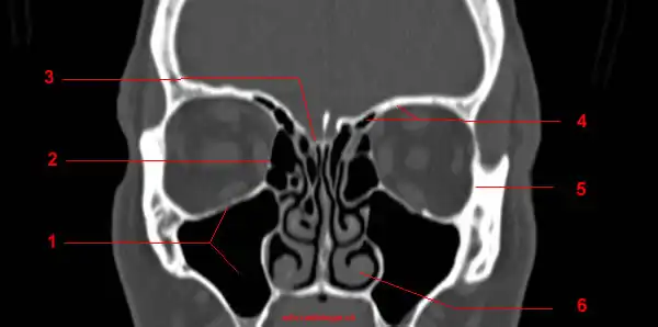
CT of the Orbit: anatomy - W-Radiology

Imaging in Skull base

Skull | Radiology Reference Article | Radiopaedia.org

Skull Base–related Lesions at Routine Head CT from the Emergency  Department: Pearls, Pitfalls, and Lessons Learned | RadioGraphics
Skull Base–related Lesions at Routine Head CT from the Emergency Department: Pearls, Pitfalls, and Lessons Learned | RadioGraphics