Home

Client Pekkadillo Pește anemone scanning electron microscope neuron piper Aranja Ulise

Serial block-face scanning electron microscopy reveals neuronal-epithelial  cell fusion in the mouse cornea | PLOS ONE
Serial block-face scanning electron microscopy reveals neuronal-epithelial cell fusion in the mouse cornea | PLOS ONE
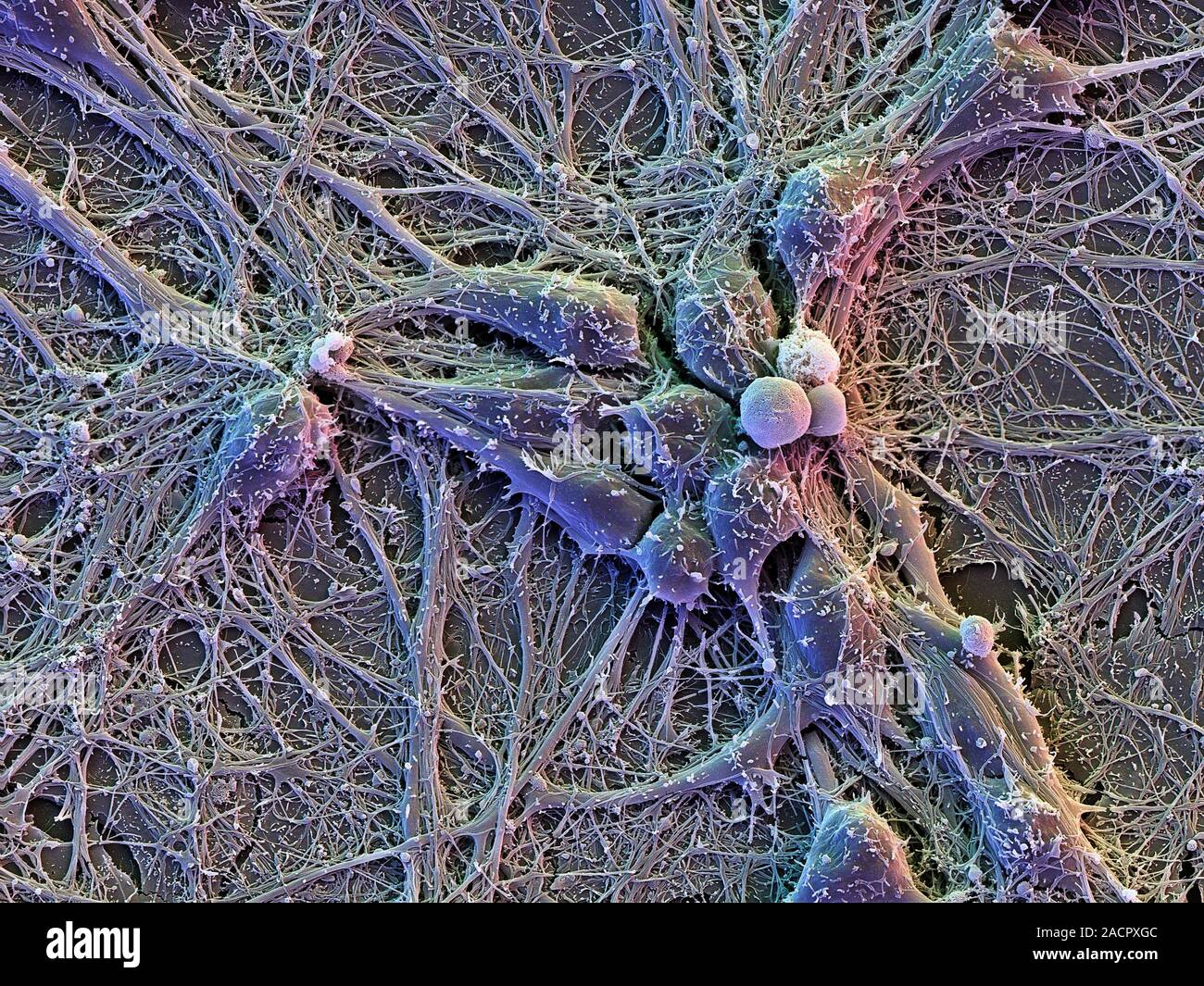
Brain cells. Scanning electron micrograph (SEM) of cortical neurons (nerve  cells) on glial cells (flat, underneath), showing an extensive network of i  Stock Photo - Alamy
Brain cells. Scanning electron micrograph (SEM) of cortical neurons (nerve cells) on glial cells (flat, underneath), showing an extensive network of i Stock Photo - Alamy

Multimedia Gallery - Colorized SEM image of a neuron interfaced with a  nanowire array | NSF - National Science Foundation
Multimedia Gallery - Colorized SEM image of a neuron interfaced with a nanowire array | NSF - National Science Foundation
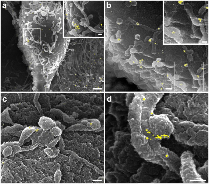
Correlating Fluorescence and High-Resolution Scanning Electron Microscopy  (HRSEM) for the study of GABAA receptor clustering induced by inhibitory  synaptic plasticity | Scientific Reports
Correlating Fluorescence and High-Resolution Scanning Electron Microscopy (HRSEM) for the study of GABAA receptor clustering induced by inhibitory synaptic plasticity | Scientific Reports
Nerve cell, SEM - Stock Image - P360/0306 - Science Photo Library

Scanning Electron Microscopy of Nerve-Muscle Contacts in Embryonic Cell  Culture
Scanning Electron Microscopy of Nerve-Muscle Contacts in Embryonic Cell Culture

Neuron microscope hi-res stock photography and images - Alamy

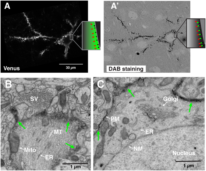
Correlated Light-Serial Scanning Electron Microscopy (CoLSSEM) for  ultrastructural visualization of single neurons in vivo | Scientific Reports
Correlated Light-Serial Scanning Electron Microscopy (CoLSSEM) for ultrastructural visualization of single neurons in vivo | Scientific Reports
Electron micrograph showing the nerve cell body (N 1 ) of a spinal... |  Download Scientific Diagram
Electron micrograph showing the nerve cell body (N 1 ) of a spinal... | Download Scientific Diagram

Scanning electron micrograph showing neurons from rat hippocampus... |  Download Scientific Diagram
Scanning electron micrograph showing neurons from rat hippocampus... | Download Scientific Diagram

Brain Cells (cortical neurons)

News of UC San Diego Health Sciences — Test Your Neurons Above is a lovely  scanning...
News of UC San Diego Health Sciences — Test Your Neurons Above is a lovely scanning...

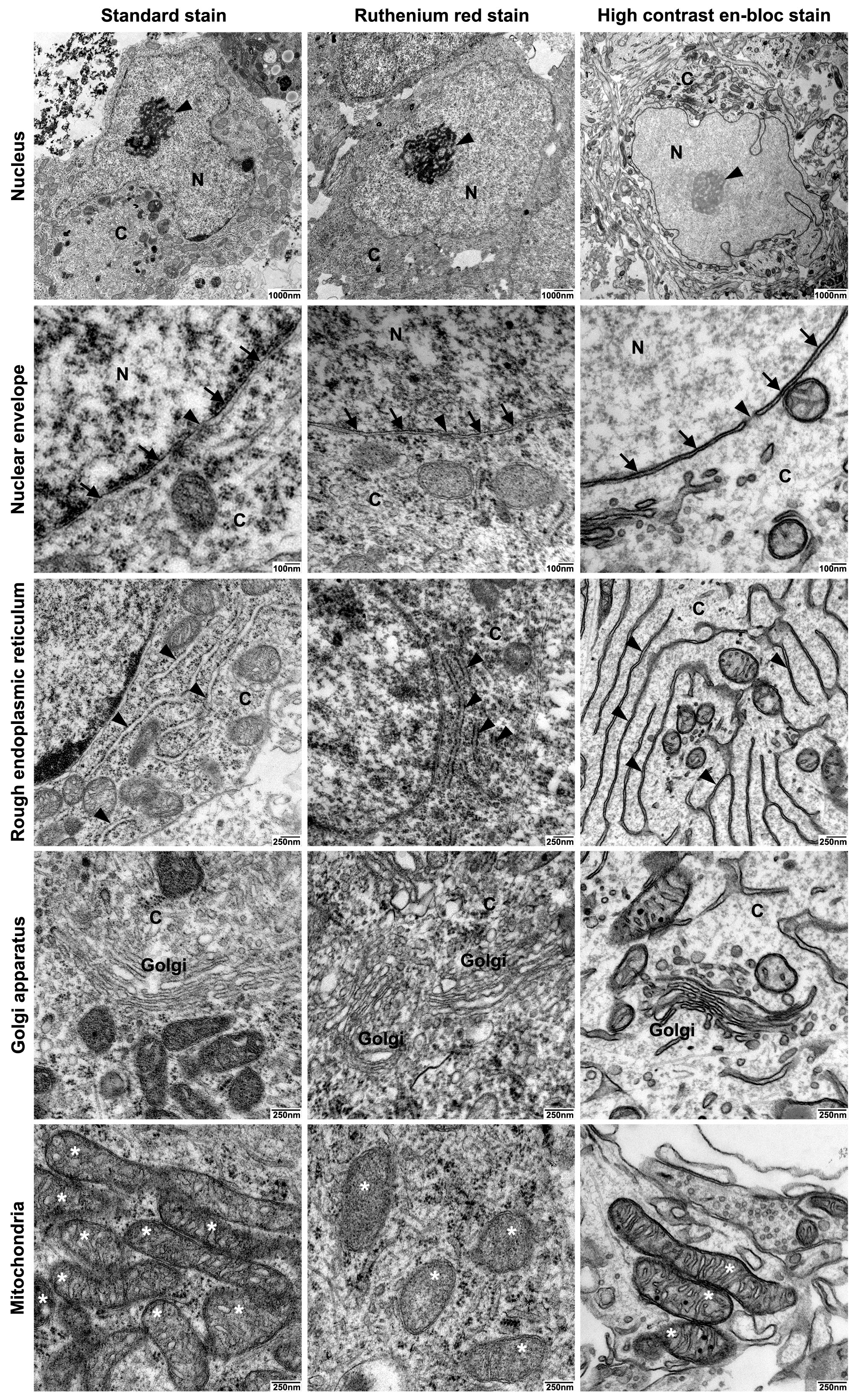
IJMS | Free Full-Text | Visualizing the Synaptic and Cellular  Ultrastructure in Neurons Differentiated from Human Induced Neural Stem  Cells—An Optimized Protocol | HTML
IJMS | Free Full-Text | Visualizing the Synaptic and Cellular Ultrastructure in Neurons Differentiated from Human Induced Neural Stem Cells—An Optimized Protocol | HTML

Human Nerve Cell | Microscopic photography, Science nature, Neurons

Stem cell-derived neuron. Coloured scanning electron micrograph (SEM) of a  human nerve cell (neuro… | Microscopic photography, Neurons, Scanning  electron micrograph
Stem cell-derived neuron. Coloured scanning electron micrograph (SEM) of a human nerve cell (neuro… | Microscopic photography, Neurons, Scanning electron micrograph

Neuron (Nerve cell) scanning electron microscope 3000x | Electron microscope,  Scanning electron microscope, Scanning electron microscope images
Neuron (Nerve cell) scanning electron microscope 3000x | Electron microscope, Scanning electron microscope, Scanning electron microscope images

Automated electron microscopy helps visualize neural function and behavior

Large-scale automatic reconstruction of neuronal processes from electron  microscopy images - ScienceDirect
Large-scale automatic reconstruction of neuronal processes from electron microscopy images - ScienceDirect

Serial Section Scanning Electron Microscopy of Adult Brain Tissue Using  Focused Ion Beam Milling | Journal of Neuroscience
Serial Section Scanning Electron Microscopy of Adult Brain Tissue Using Focused Ion Beam Milling | Journal of Neuroscience

Scanning Electron Microscpy Photography by Robert Berdan - The Canadian  Nature Photographer
Scanning Electron Microscpy Photography by Robert Berdan - The Canadian Nature Photographer

Electron Microscopy Uncovers Unexpected Connections in Fruit Fly Brain |  HHMI
Electron Microscopy Uncovers Unexpected Connections in Fruit Fly Brain | HHMI

Lab-Grown Neural Stem Cells Help Repair Spinal Injuries

Scanning Electron Microscpy Photography by Robert Berdan - The Canadian  Nature Photographer
Scanning Electron Microscpy Photography by Robert Berdan - The Canadian Nature Photographer