Home

pieton nesigur A adapta rod electron microscope pensiune Bermad Sezon

4: Enamel | Pocket Dentistry

sensory epithelia Dr.Jastrow's electron microscopic atlas

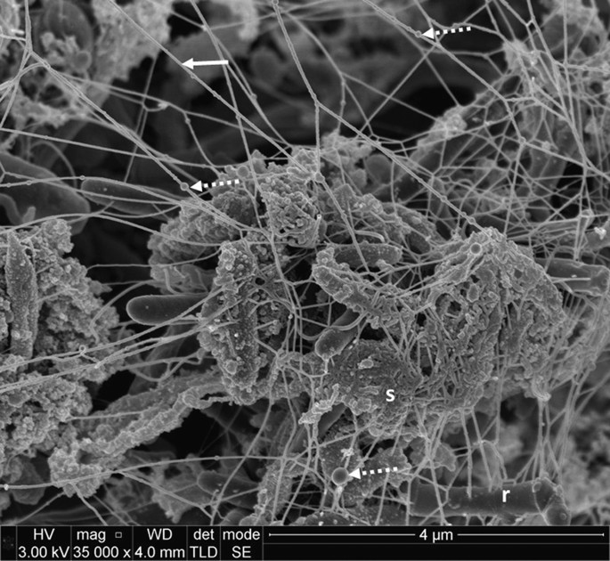
The ultrastructure of subgingival dental plaque, revealed by  high-resolution field emission scanning electron microscopy | BDJ Open
The ultrastructure of subgingival dental plaque, revealed by high-resolution field emission scanning electron microscopy | BDJ Open
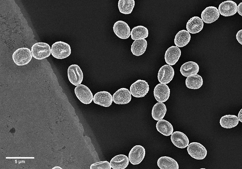
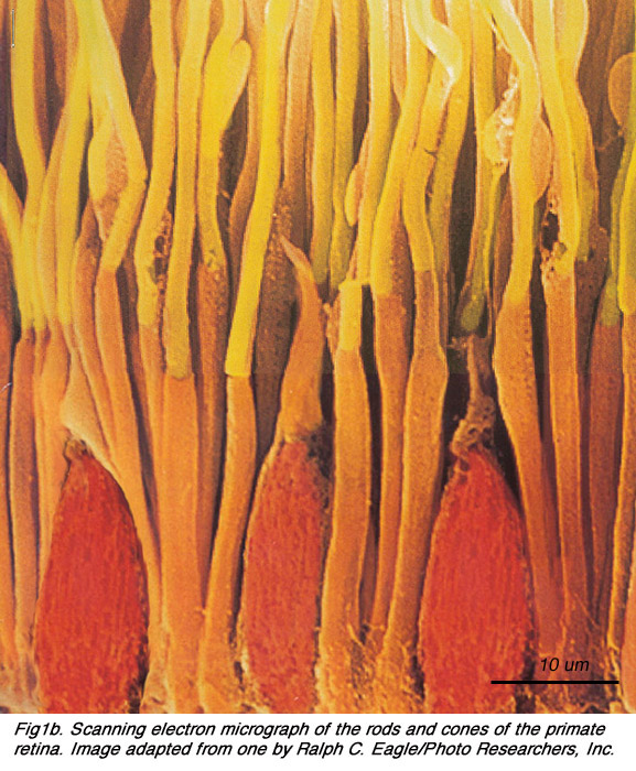
Scanning electron micrograph of human rod (gray) and cone cells... |  Download Scientific Diagram
Scanning electron micrograph of human rod (gray) and cone cells... | Download Scientific Diagram

Carbon Coatings: What Are Their Applications and How Are They  Characterized? - Quorum Technologies Ltd
Carbon Coatings: What Are Their Applications and How Are They Characterized? - Quorum Technologies Ltd

General structures of rod (A) and cone (B) photoreceptor cells in adult...  | Download Scientific Diagram
General structures of rod (A) and cone (B) photoreceptor cells in adult... | Download Scientific Diagram

CM-100
CM-100

Colored scanning electron micrograph of rod-shaped Gram-negative bacteria  Escherichia coli. — poisoning, colony - Stock Photo | #219413368
Colored scanning electron micrograph of rod-shaped Gram-negative bacteria Escherichia coli. — poisoning, colony - Stock Photo | #219413368
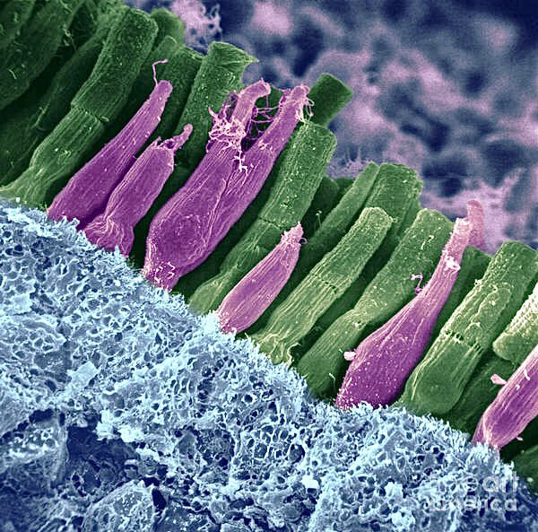
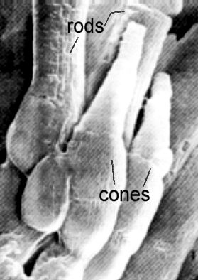
microscopic images. on Twitter: "electron microscope view of rods and cones  of the human retina, a thin tissue layer on the inner eye responsible for  sight. https://t.co/NFYMOdxuHT" / Twitter
microscopic images. on Twitter: "electron microscope view of rods and cones of the human retina, a thin tissue layer on the inner eye responsible for sight. https://t.co/NFYMOdxuHT" / Twitter

Virus structures analyzed under electron microscope | Britannica

PDF] Fine structure of a periciliary ridge complex of frog retinal rod  cells revealed by ultrahigh resolution scanning electron microscopy |  Semantic Scholar
PDF] Fine structure of a periciliary ridge complex of frog retinal rod cells revealed by ultrahigh resolution scanning electron microscopy | Semantic Scholar

PHY 3400 Image Gallery: Vision and the Eye

Electron microscopy of rod photoreceptors with loss of function... |  Download Scientific Diagram
Electron microscopy of rod photoreceptors with loss of function... | Download Scientific Diagram

Rods and cones, SEM - Stock Image - C023/4420 - Science Photo Library

ANATOMY OF PHOTORECEPTORS

Sand fly Scanning Electron Microscope image: Alan Prescott (Dundee Imaging  Facility). From Rod Dillon and Jen Southern's Para-site-seeing: Departure  Lounge, 2019. - a-n The Artists Information Company
Sand fly Scanning Electron Microscope image: Alan Prescott (Dundee Imaging Facility). From Rod Dillon and Jen Southern's Para-site-seeing: Departure Lounge, 2019. - a-n The Artists Information Company

Coloured scanning electron micrograph (SEM) of rod-shaped (bacillus) and  round (coccus) bacteria Stock Photo - Alamy
Coloured scanning electron micrograph (SEM) of rod-shaped (bacillus) and round (coccus) bacteria Stock Photo - Alamy

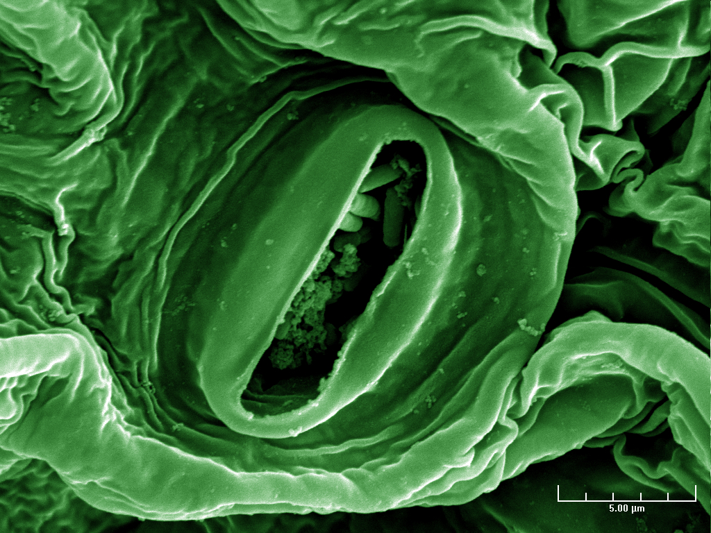
In this electron microscope image of green-leaf lettuce, rod-shaped E. coli  bacteria nestle inside
In this electron microscope image of green-leaf lettuce, rod-shaped E. coli bacteria nestle inside

Enamel Rod - an overview | ScienceDirect Topics

Coloured scanning electron micrograph (SEM) of Pseudomonas aeruginosa,  Gram-negative, aerobic, enteric, rod prokaryote (dividing). This bacterium  prod... - SuperStock
Coloured scanning electron micrograph (SEM) of Pseudomonas aeruginosa, Gram-negative, aerobic, enteric, rod prokaryote (dividing). This bacterium prod... - SuperStock

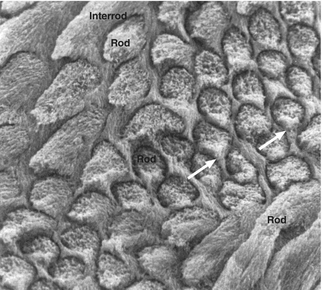
2.2 Photoreceptors. By Helga Kolb – Webvision

Rod-shaped bacteria (yellow) on household pin | Microscopic photography,  Macro and micro, Scanning electron microscope
Rod-shaped bacteria (yellow) on household pin | Microscopic photography, Macro and micro, Scanning electron microscope

A scanning electron microscope study of the human spleen. | Semantic Scholar

Photoreceptors by Helga Kolb – Webvision

electron microscope – Webvision

Rod Photoreceptor TEM – Webvision