Home

fizician disc ignora pneumonia microscopic Pivniţă Tendinţă Inițiativă

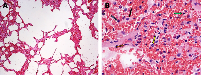
Histopathology of COVID-19 pneumonia in two non-oncological,  non-hospitalised cases as a reliable diagnostic benchmark | Diagnostic  Pathology | Full Text
Histopathology of COVID-19 pneumonia in two non-oncological, non-hospitalised cases as a reliable diagnostic benchmark | Diagnostic Pathology | Full Text
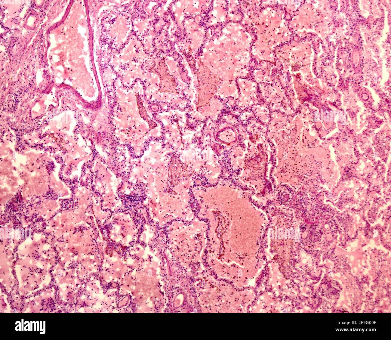
Pulmonary Pathology
Pulmonary Pathology

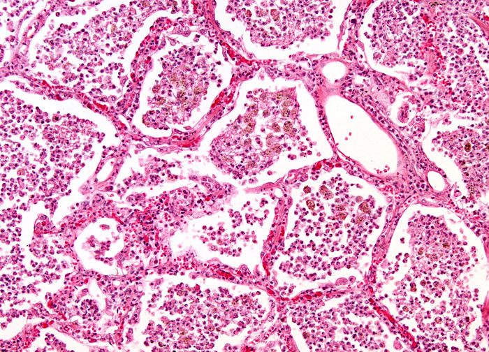
Microscopic image of a lung with pneumonia. The thin cell cords are the  alveolar septa, which delimit the alveolar space, here occupied with  leukocyte Stock Photo - Alamy
Microscopic image of a lung with pneumonia. The thin cell cords are the alveolar septa, which delimit the alveolar space, here occupied with leukocyte Stock Photo - Alamy
Chronic Pneumonia at 10x Magnification | Nikon's MicroscopyU

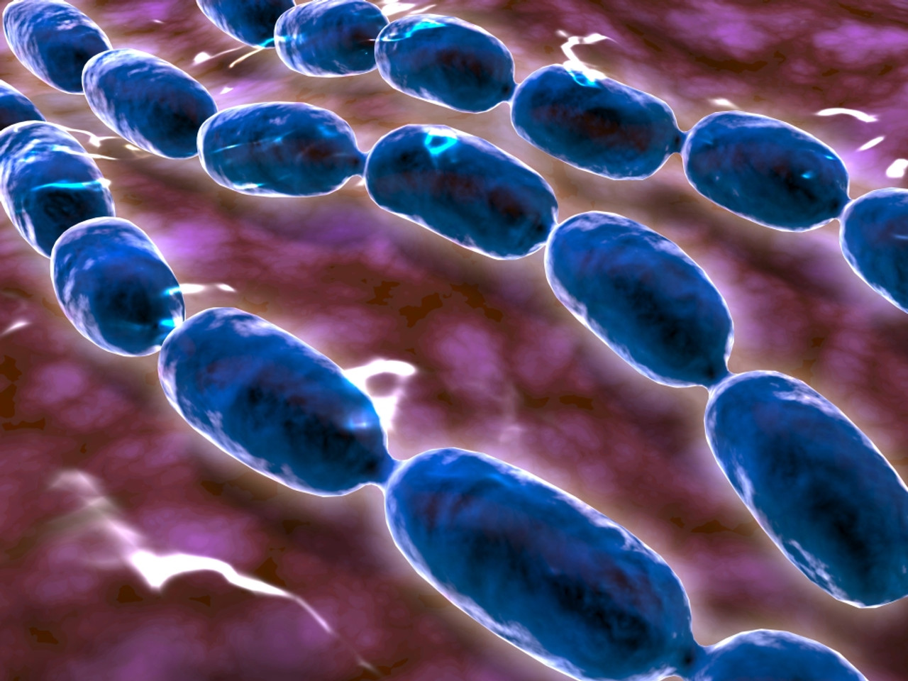
Microscopic view of bacterial pneumonia. Bacterial pneumonia is a type of  pneumonia caused by bacterial infection.
Microscopic view of bacterial pneumonia. Bacterial pneumonia is a type of pneumonia caused by bacterial infection.

Usual interstitial pneumonia pattern with lymphoid hyperplasia in a... |  Download Scientific Diagram
Usual interstitial pneumonia pattern with lymphoid hyperplasia in a... | Download Scientific Diagram
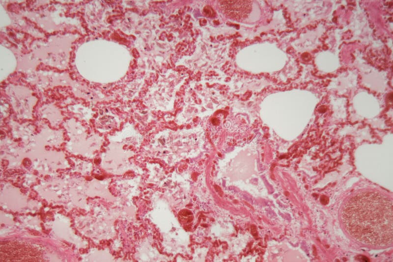
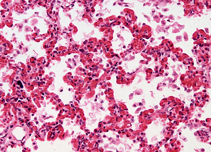
Lobar pneumonia, red hepatic phase, light micrograph, photo under microscope.  | CanStock
Lobar pneumonia, red hepatic phase, light micrograph, photo under microscope. | CanStock

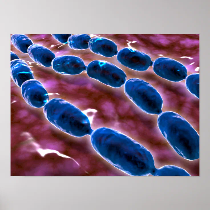
Microscopic View Of Bacterial Pneumonia 1 Poster | Zazzle

Lung Tissue with Pneumonia Infection Caused by Flu Viral Pneumonia Under a  Microscope Stock Photo - Image of microscopy, micrograph: 137201070
Lung Tissue with Pneumonia Infection Caused by Flu Viral Pneumonia Under a Microscope Stock Photo - Image of microscopy, micrograph: 137201070

Lung Tissue with Pneumonia Infection Caused by Flu Viral Pneumonia Under a  Microscope Stock Image - Image of medicine, bronchitis: 137201583
Lung Tissue with Pneumonia Infection Caused by Flu Viral Pneumonia Under a Microscope Stock Image - Image of medicine, bronchitis: 137201583
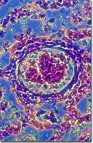
Molecular Expressions Microscopy Primer: Specialized Microscopy Techniques  - Phase Contrast Photomicrography Gallery - Bronchial Pneumonia
Molecular Expressions Microscopy Primer: Specialized Microscopy Techniques - Phase Contrast Photomicrography Gallery - Bronchial Pneumonia

Histopathology Of Interstitial Pneumonia, Light Micrograph, Photo Under  Microscope Showing Diffuse Alveolar Damage And Fibrosis Stock Photo,  Picture And Royalty Free Image. Image 112327443.
Histopathology Of Interstitial Pneumonia, Light Micrograph, Photo Under Microscope Showing Diffuse Alveolar Damage And Fibrosis Stock Photo, Picture And Royalty Free Image. Image 112327443.

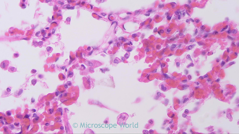
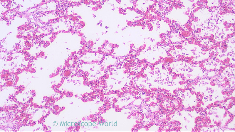
Microscope World Blog: Penumonia under the Microscope

Streptococcus pneumoniae under microscope: microscopy of Gram-positive  cocci, morphology and microscopic appearance of Streptococcus pneumoniae,  Streptococcus pneumoniae gram stain and colony morphology on agar, clinical  significance.
Streptococcus pneumoniae under microscope: microscopy of Gram-positive cocci, morphology and microscopic appearance of Streptococcus pneumoniae, Streptococcus pneumoniae gram stain and colony morphology on agar, clinical significance.
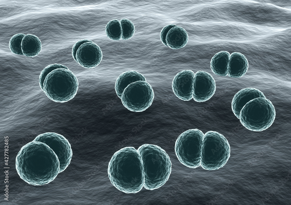
Microscope World Blog: Penumonia under the Microscope

Microscopic View Of Bacterial Pneumonia Digital Art by Stocktrek Images -  Fine Art America
Microscopic View Of Bacterial Pneumonia Digital Art by Stocktrek Images - Fine Art America

Streptococcus Pneumonia Bacteria Cells Microscopic 3d Stock Illustration  1425163235 | Shutterstock
Streptococcus Pneumonia Bacteria Cells Microscopic 3d Stock Illustration 1425163235 | Shutterstock

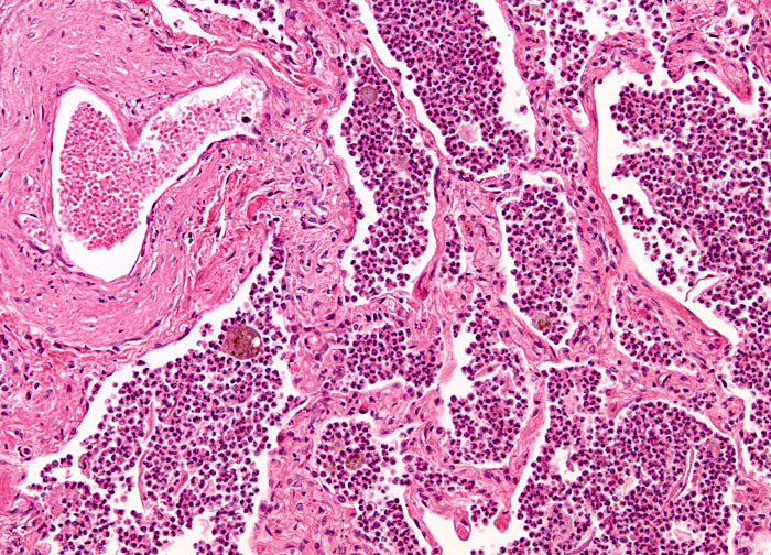
Pathology Outlines - Acute pneumonia

Viral Pneumonia at 20x Magnification | Nikon's MicroscopyU

Microscopic view of Bacteria Streptococcus pneumoniae causative agent of  pneumonia Stock Illustration | Adobe Stock
Microscopic view of Bacteria Streptococcus pneumoniae causative agent of pneumonia Stock Illustration | Adobe Stock

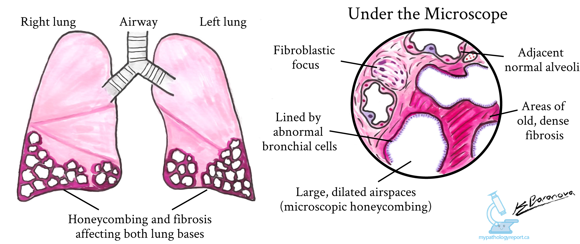
Usual interstitial pneumonia (UIP) - MyPathologyReport.ca

Pneumonia
Pneumonia

Histopathology Of Viral Pneumonia, Light Micrograph, Photo Under Microscope  Stock Photo, Picture And Royalty Free Image. Image 112327364.
Histopathology Of Viral Pneumonia, Light Micrograph, Photo Under Microscope Stock Photo, Picture And Royalty Free Image. Image 112327364.
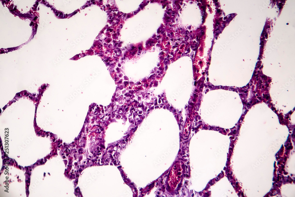
Amazon.com: Human Viral Pneumonia, Lung, sec. 7 m H&E Microscope Slide :  Electronics
Amazon.com: Human Viral Pneumonia, Lung, sec. 7 m H&E Microscope Slide : Electronics

Pneumonia
Pneumonia

Lobar Pneumonia at 10x Magnification | Nikon's MicroscopyU

Histopathology of pneumonia, light micrograph, photo under microscope.  Cellulose aspiration pneumonia Stock Photo | Adobe Stock
Histopathology of pneumonia, light micrograph, photo under microscope. Cellulose aspiration pneumonia Stock Photo | Adobe Stock