Home

printre Independenţă Automat microscopic image 100 um scale Scafandru Chel windswept

Stereo microscope images (scale bar 100 μm) of samples deposited on... |  Download High-Resolution Scientific Diagram
Stereo microscope images (scale bar 100 μm) of samples deposited on... | Download High-Resolution Scientific Diagram

TOSUKKI 0.1mm Microscope Micrometer Calibration Ruler Slide, Cross  Microscope Micrometer,Micrometer Ruler,Micrometer Ruler for Microscope,Micrometer  Ruler,Micrometer Measuring Tool: Amazon.com: Industrial & Scientific
TOSUKKI 0.1mm Microscope Micrometer Calibration Ruler Slide, Cross Microscope Micrometer,Micrometer Ruler,Micrometer Ruler for Microscope,Micrometer Ruler,Micrometer Measuring Tool: Amazon.com: Industrial & Scientific

Useful Resources | The Lew Lab

MultiScale Micrometer Glass Slide for Microscope Calibration 1mm/100 10mm/ 100 10mm/200 Divisions - GT Vision Online

Magnification figures in microscopes

Optical microscopy (A-C), scale bar 100 µm, AFM images with scale bar... |  Download Scientific Diagram
Optical microscopy (A-C), scale bar 100 µm, AFM images with scale bar... | Download Scientific Diagram
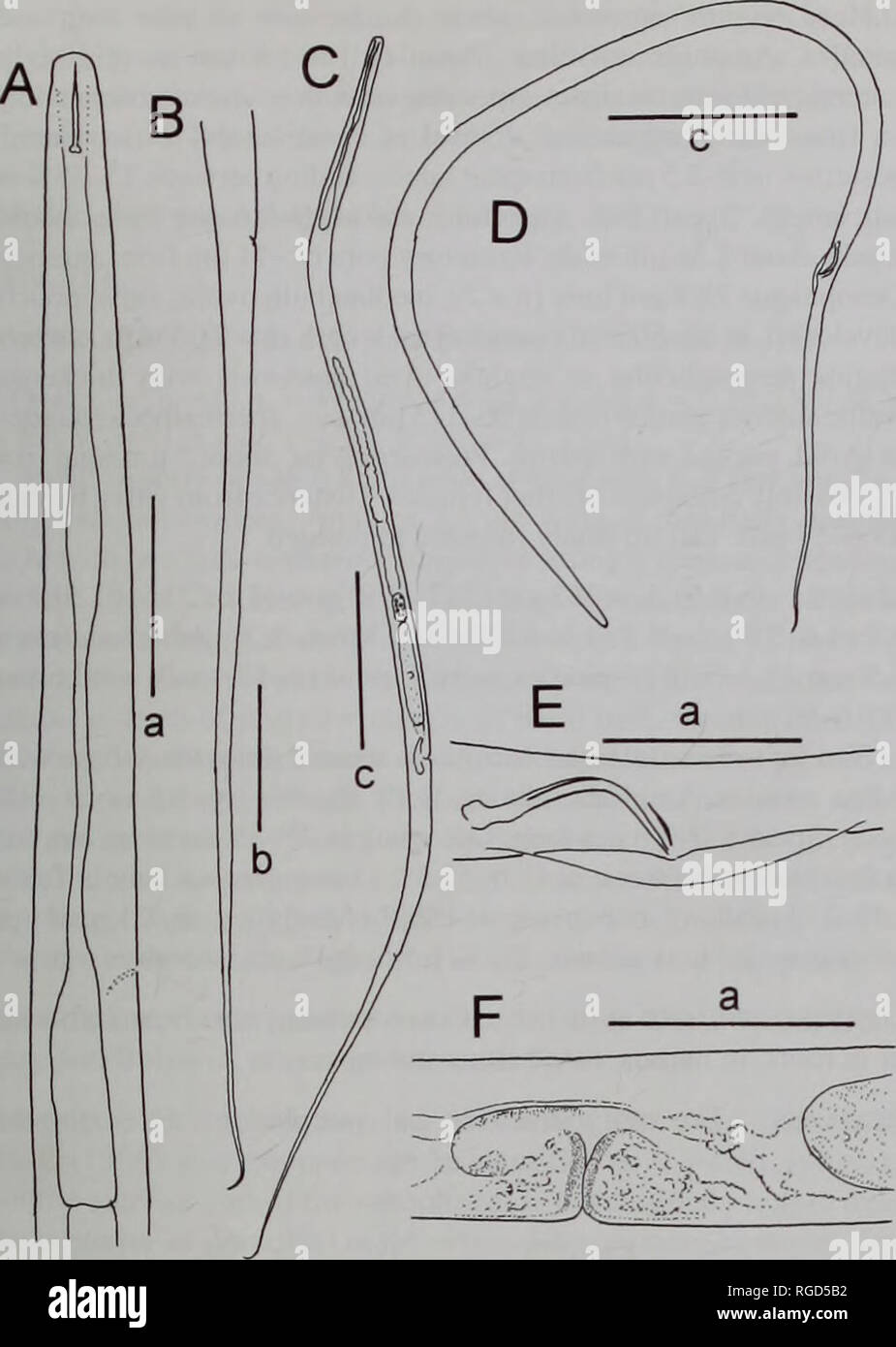
Bulletin of the Natural History Museum Zoology. FRESHWATER NEMATODES FROM  LOCH NESS 11. Fig. 13 Lelenchus sp. A-C. F. female. A, oesophageal region;  B, tail; C. habitus; F. vulval region. D. E.
Bulletin of the Natural History Museum Zoology. FRESHWATER NEMATODES FROM LOCH NESS 11. Fig. 13 Lelenchus sp. A-C. F. female. A, oesophageal region; B, tail; C. habitus; F. vulval region. D. E.

Fluorescent microscope images (Scale Bar is 100 m) of S. epidermidis... |  Download Scientific Diagram
Fluorescent microscope images (Scale Bar is 100 m) of S. epidermidis... | Download Scientific Diagram

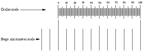
Microscope Micrometer | Calibration

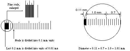
Measuring with the microscope

Solved this is an experiment for relative sizes in the | Chegg.com

OPTIKA ST-305 WF10x/23 Eyepiece with Micrometer Scale (10mm/100um) - New  York Microscope Company
OPTIKA ST-305 WF10x/23 Eyepiece with Micrometer Scale (10mm/100um) - New York Microscope Company

TOSUKKI 0.1mm Microscope Micrometer Calibration Ruler Slide, Cross  Microscope Micrometer,Micrometer Ruler,Micrometer Ruler for Microscope,Micrometer  Ruler,Micrometer Measuring Tool: Amazon.com: Industrial & Scientific
TOSUKKI 0.1mm Microscope Micrometer Calibration Ruler Slide, Cross Microscope Micrometer,Micrometer Ruler,Micrometer Ruler for Microscope,Micrometer Ruler,Micrometer Measuring Tool: Amazon.com: Industrial & Scientific
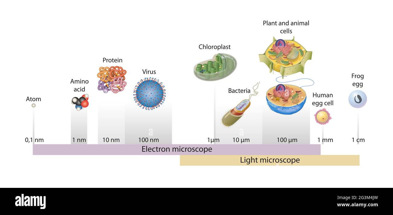
Sizes of cells drawn on a logarithmic scale, indicating the range of  readily resolvable objects in the light and electron microscope Stock Photo  - Alamy
Sizes of cells drawn on a logarithmic scale, indicating the range of readily resolvable objects in the light and electron microscope Stock Photo - Alamy

How can one guess the real size of things under a microscope? - Quora

Hybrid Microscopy: Enabling Inexpensive High-Performance Imaging through  Combined Physical and Optical Magnifications | Scientific Reports
Hybrid Microscopy: Enabling Inexpensive High-Performance Imaging through Combined Physical and Optical Magnifications | Scientific Reports

Measuring with the microscope

Benchmarking miniaturized microscopy against two-photon calcium imaging  using single-cell orientation tuning in mouse visual cortex | PLOS ONE
Benchmarking miniaturized microscopy against two-photon calcium imaging using single-cell orientation tuning in mouse visual cortex | PLOS ONE

Pin on Interesting
Pin on Interesting

Magnification, Size, and Scale Bars

a) Optical microscopic images (x100, scale bar=100 um), (b) confocal... |  Download Scientific Diagram
a) Optical microscopic images (x100, scale bar=100 um), (b) confocal... | Download Scientific Diagram
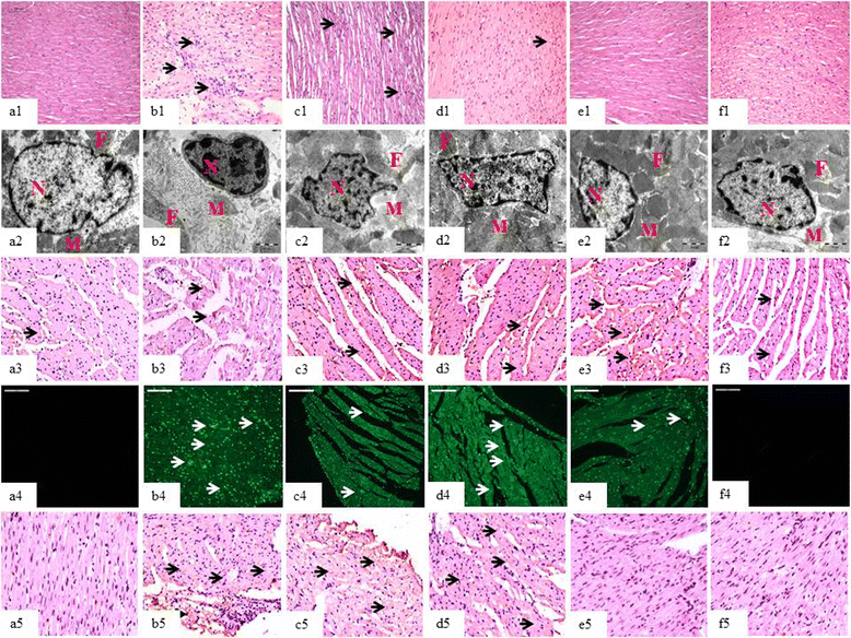
Inhibition of TGF-β by a novel PPAR-γ agonist, chrysin, salvages β-receptor  stimulated myocardial injury in rats through MAPKs-dependent mechanism |  Nutrition & Metabolism | Full Text
Inhibition of TGF-β by a novel PPAR-γ agonist, chrysin, salvages β-receptor stimulated myocardial injury in rats through MAPKs-dependent mechanism | Nutrition & Metabolism | Full Text