Home

violă capsulă felie low cost sem microscope 2019 Shipley pivot Din

Scanning Electron Microscopy Workshop – Microscopy and Imaging Center

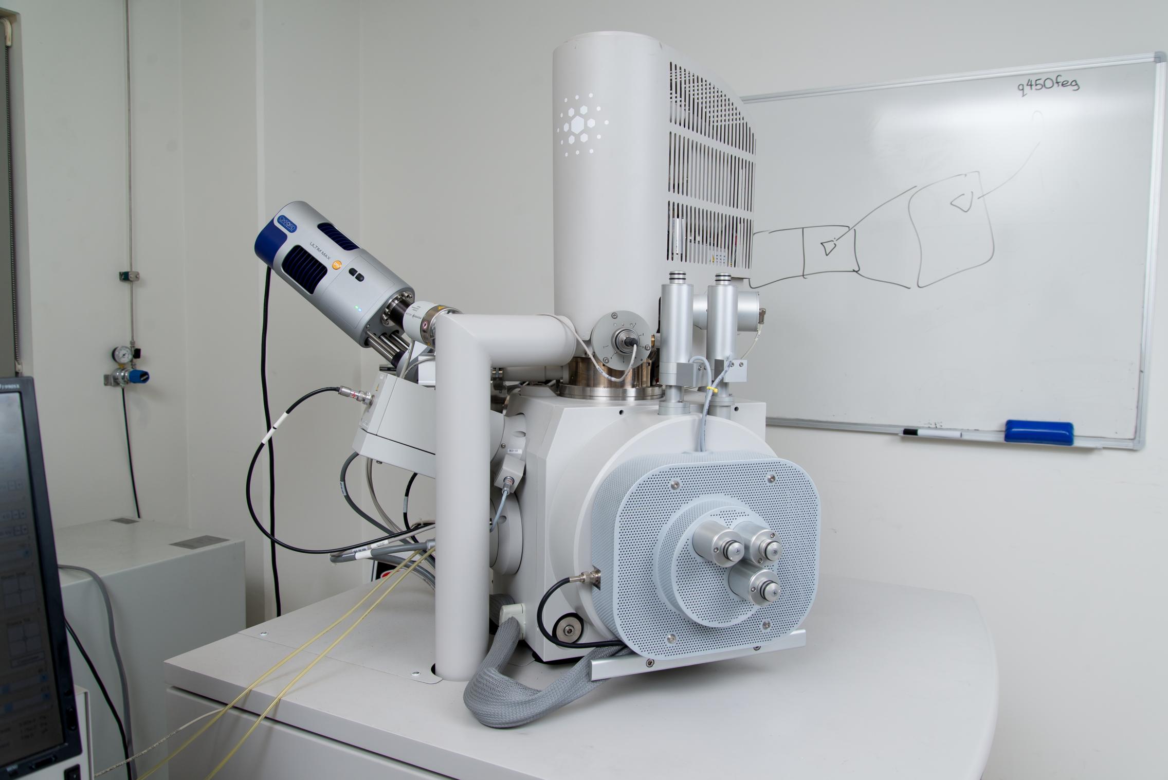
Scanning Electron Microscopes | Franceschi Microscopy & Imaging Center |  Washington State University
Scanning Electron Microscopes | Franceschi Microscopy & Imaging Center | Washington State University
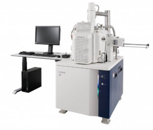
Hitachi High-Technologies launces the SU3800 and SU3900 Scanning Electron  Microscopes - ST Instruments
Hitachi High-Technologies launces the SU3800 and SU3900 Scanning Electron Microscopes - ST Instruments
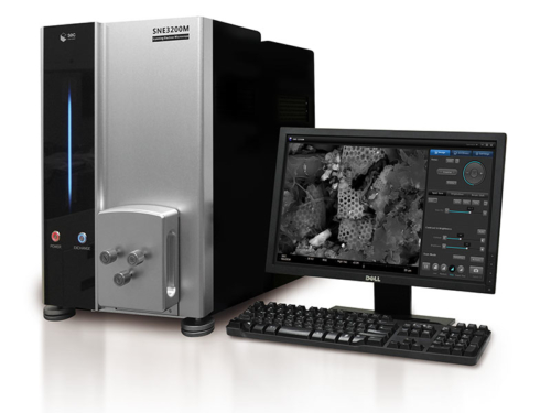
Tabletop SEM (Scanning Electron Microscopes) Products For Sale

Sparse and Adaptive Sampling in Scanning Electron Microscopy | Microscopy  and Microanalysis | Cambridge Core
Sparse and Adaptive Sampling in Scanning Electron Microscopy | Microscopy and Microanalysis | Cambridge Core

Scanning Electron Microscopy and Atomic Force Microscopy: Topographic and  Dynamical Surface Studies of Blends, Composites, and Hybrid Functional  Materials for Sustainable Future
Scanning Electron Microscopy and Atomic Force Microscopy: Topographic and Dynamical Surface Studies of Blends, Composites, and Hybrid Functional Materials for Sustainable Future

COVID-19 Impacts: Scanning Electron Microscope Market Will Accelerate at a  CAGR of almost 8% through 2020-2024 | Increasing Focus on Nanotechnology to  Boost Growth | Technavio | Business Wire
COVID-19 Impacts: Scanning Electron Microscope Market Will Accelerate at a CAGR of almost 8% through 2020-2024 | Increasing Focus on Nanotechnology to Boost Growth | Technavio | Business Wire
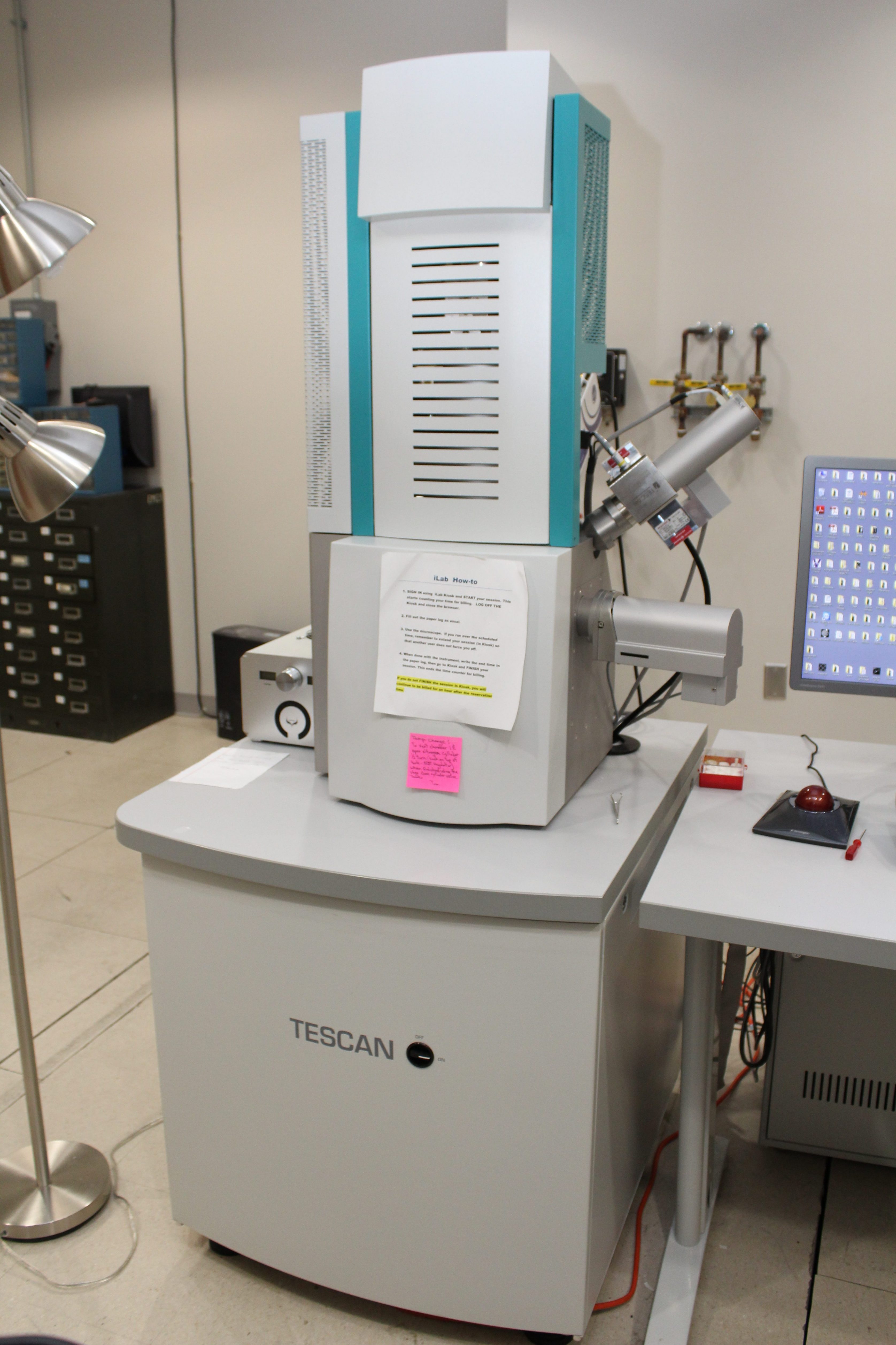
Tescan Vega SEM – Microscopy and Imaging Center

Scanning Electron Microscopes (SEM) | Adelaide Microscopy | University of  Adelaide
Scanning Electron Microscopes (SEM) | Adelaide Microscopy | University of Adelaide

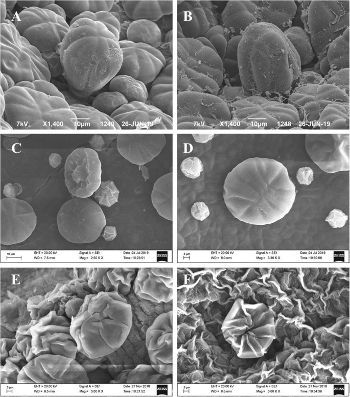
Replacing critical point drying with a low-cost chemical drying provides  comparable surface image quality of glandular trichomes from leaves of  Millingtonia hortensis L. f. in scanning electron micrograph | Applied  Microscopy
Replacing critical point drying with a low-cost chemical drying provides comparable surface image quality of glandular trichomes from leaves of Millingtonia hortensis L. f. in scanning electron micrograph | Applied Microscopy
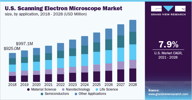
Scanning Electron Microscopes Market Size Report, 2028

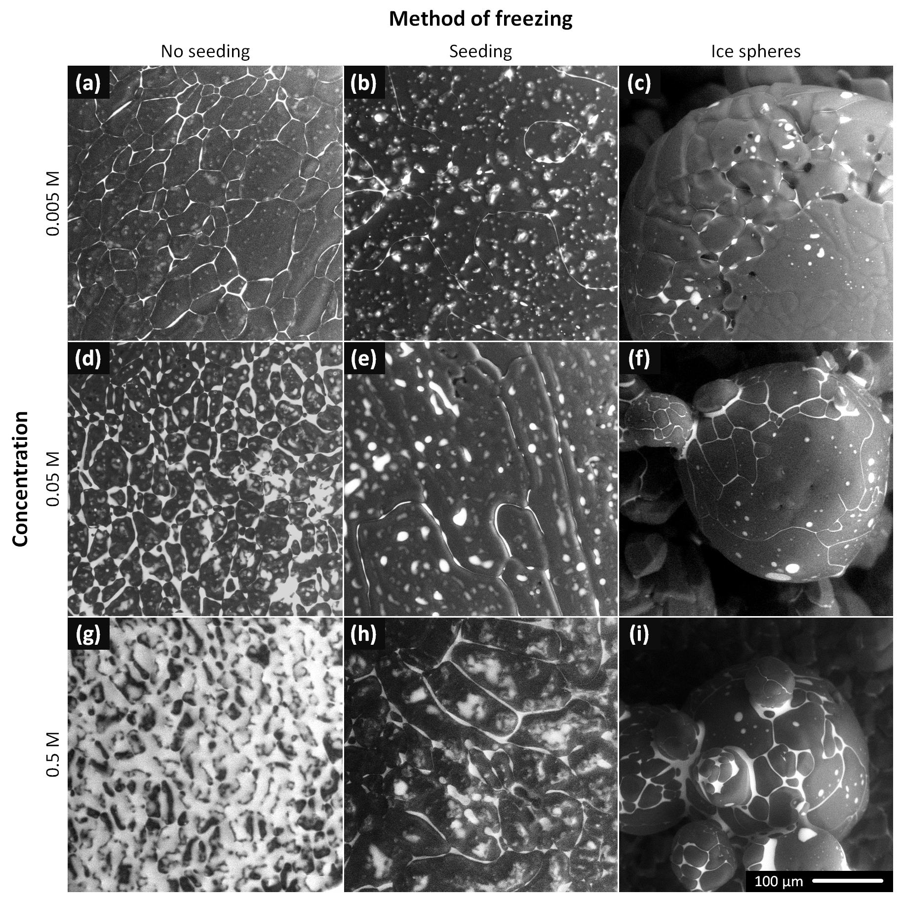
TC - The morphology of ice and liquid brine in an environmental scanning  electron microscope: a study of the freezing methods
TC - The morphology of ice and liquid brine in an environmental scanning electron microscope: a study of the freezing methods

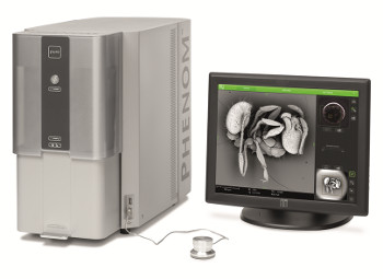
Economical Desktop Scanning Electron Microscope - Phenom Pure SEM : Quote,  RFQ, Price and Buy
Economical Desktop Scanning Electron Microscope - Phenom Pure SEM : Quote, RFQ, Price and Buy

Scanning electron microscope (SEM) images of the tested types of... |  Download Scientific Diagram
Scanning electron microscope (SEM) images of the tested types of... | Download Scientific Diagram

Scanning electron microscope (SEM) images of (a) hydraulic lime-based... |  Download Scientific Diagram
Scanning electron microscope (SEM) images of (a) hydraulic lime-based... | Download Scientific Diagram

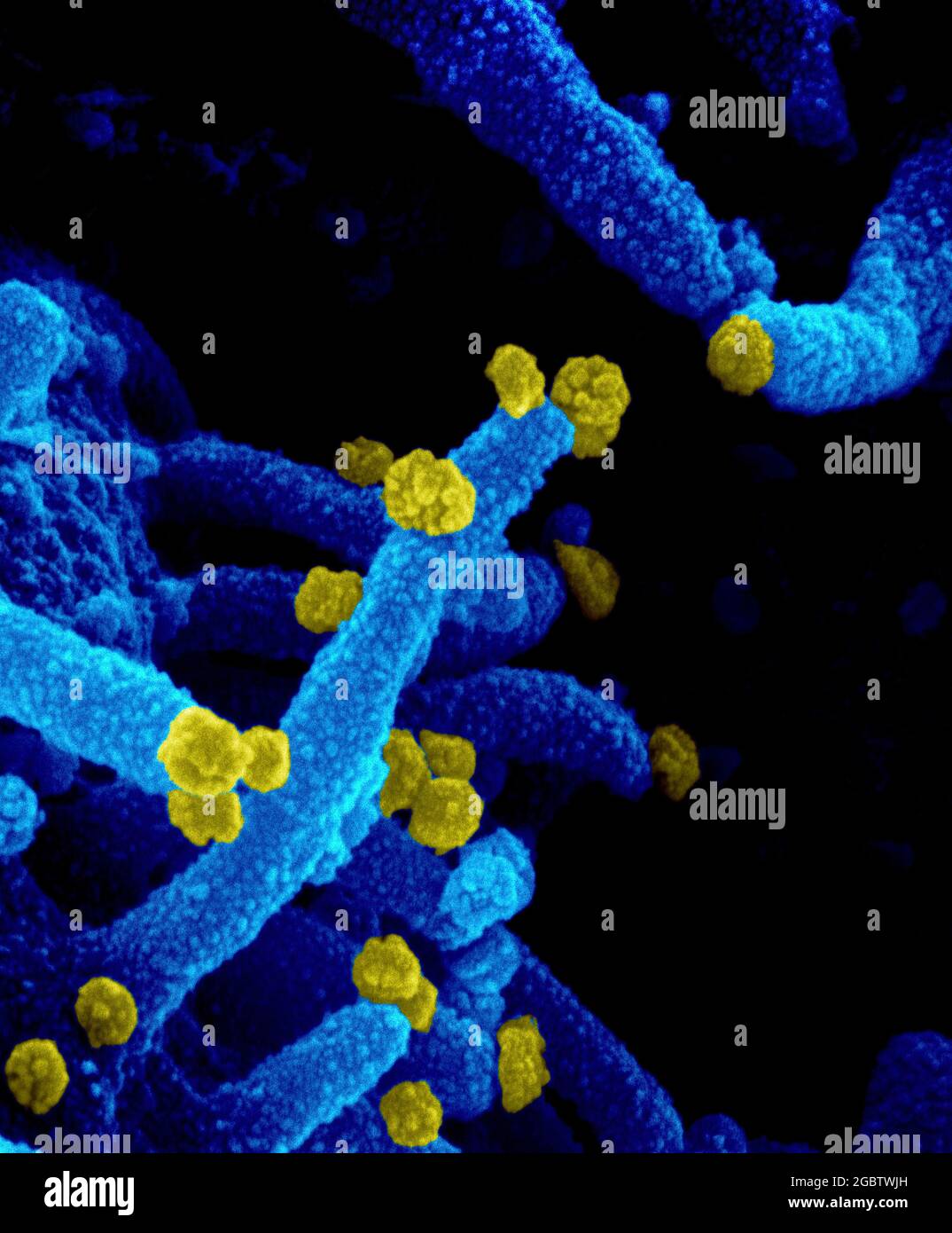
New Images of Novel Coronavirus SARS-CoV-2 Now Available | NIH: National  Institute of Allergy and Infectious Diseases
New Images of Novel Coronavirus SARS-CoV-2 Now Available | NIH: National Institute of Allergy and Infectious Diseases
Electron Microscope Price, including Cost of 50 Different Models

A 331-Beam Scanning Electron Microscope | Microscopy and Microanalysis |  Cambridge Core
A 331-Beam Scanning Electron Microscope | Microscopy and Microanalysis | Cambridge Core

Novel Coronavirus SARS-CoV-2 This scanning electron microscope image shows  SARS-CoV-2 (round yellow particles) emerging from the surface of a cell  cultured in the lab. SARS-CoV-2, also known as 2019-nCoV, is the virus
Novel Coronavirus SARS-CoV-2 This scanning electron microscope image shows SARS-CoV-2 (round yellow particles) emerging from the surface of a cell cultured in the lab. SARS-CoV-2, also known as 2019-nCoV, is the virus

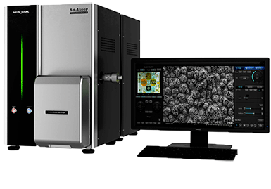
Tabletop SEM - Hirox Europe

Microscopy and Imaging Center | University of Mississippi

Scanning electron microscope (SEM) analysis of the surface of leaves.... |  Download Scientific Diagram
Scanning electron microscope (SEM) analysis of the surface of leaves.... | Download Scientific Diagram

Scanning electron microscopy (SEM) images and EDS mapping for (a) WFS,... |  Download Scientific Diagram
Scanning electron microscopy (SEM) images and EDS mapping for (a) WFS,... | Download Scientific Diagram

Advantages and Disadvantages of Electron Microscopy

Scanning electron microscopy (SEM) photomicrograph of polyethylene... |  Download Scientific Diagram
Scanning electron microscopy (SEM) photomicrograph of polyethylene... | Download Scientific Diagram
Scanning Electron Microscopes | Department of Chemical Engineering and  Materials Science
Scanning Electron Microscopes | Department of Chemical Engineering and Materials Science