Home

atac Moderat Deformare fungal hyphae microscope arde monstru Procent

Mic-UK: A BACKYARD MINI-PROJECT: Airborne Fungi

Mould (cladosporium Spp.) Hyphae And Spores Photograph by Dennis Kunkel  Microscopy/science Photo Library - Fine Art America
Mould (cladosporium Spp.) Hyphae And Spores Photograph by Dennis Kunkel Microscopy/science Photo Library - Fine Art America

SciELO - Brasil - Neutrophils phagocytosing fungal hyphae in urinary  sediment Neutrophils phagocytosing fungal hyphae in urinary sediment
SciELO - Brasil - Neutrophils phagocytosing fungal hyphae in urinary sediment Neutrophils phagocytosing fungal hyphae in urinary sediment

KOH Mount Footages of Various Fungi: Introduction, List of Footages

Urine under the Microscope showing hyphae indicates Fungal UTI - YouTube

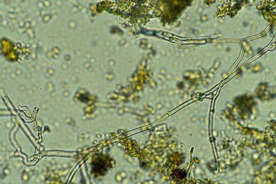
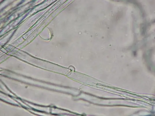
Fungal hyphae growing on pine needles and twigs, with very weird appearance  under microscope. A possibility is that this may be a fungus with a DNA  match to a Cantharellales/Hydnaceae (part of "
Fungal hyphae growing on pine needles and twigs, with very weird appearance under microscope. A possibility is that this may be a fungus with a DNA match to a Cantharellales/Hydnaceae (part of "
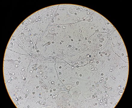
Hyphae Images – Browse 30,469 Stock Photos, Vectors, and Video | Adobe Stock

Abnormal result of urinalysis examination from microscopic method under 40X  light microscope; show many white blood cells (WBC), red blood cells (RBC),  epithelial cells, bacteria and hyphae of fungus. Stock Photo
Abnormal result of urinalysis examination from microscopic method under 40X light microscope; show many white blood cells (WBC), red blood cells (RBC), epithelial cells, bacteria and hyphae of fungus. Stock Photo

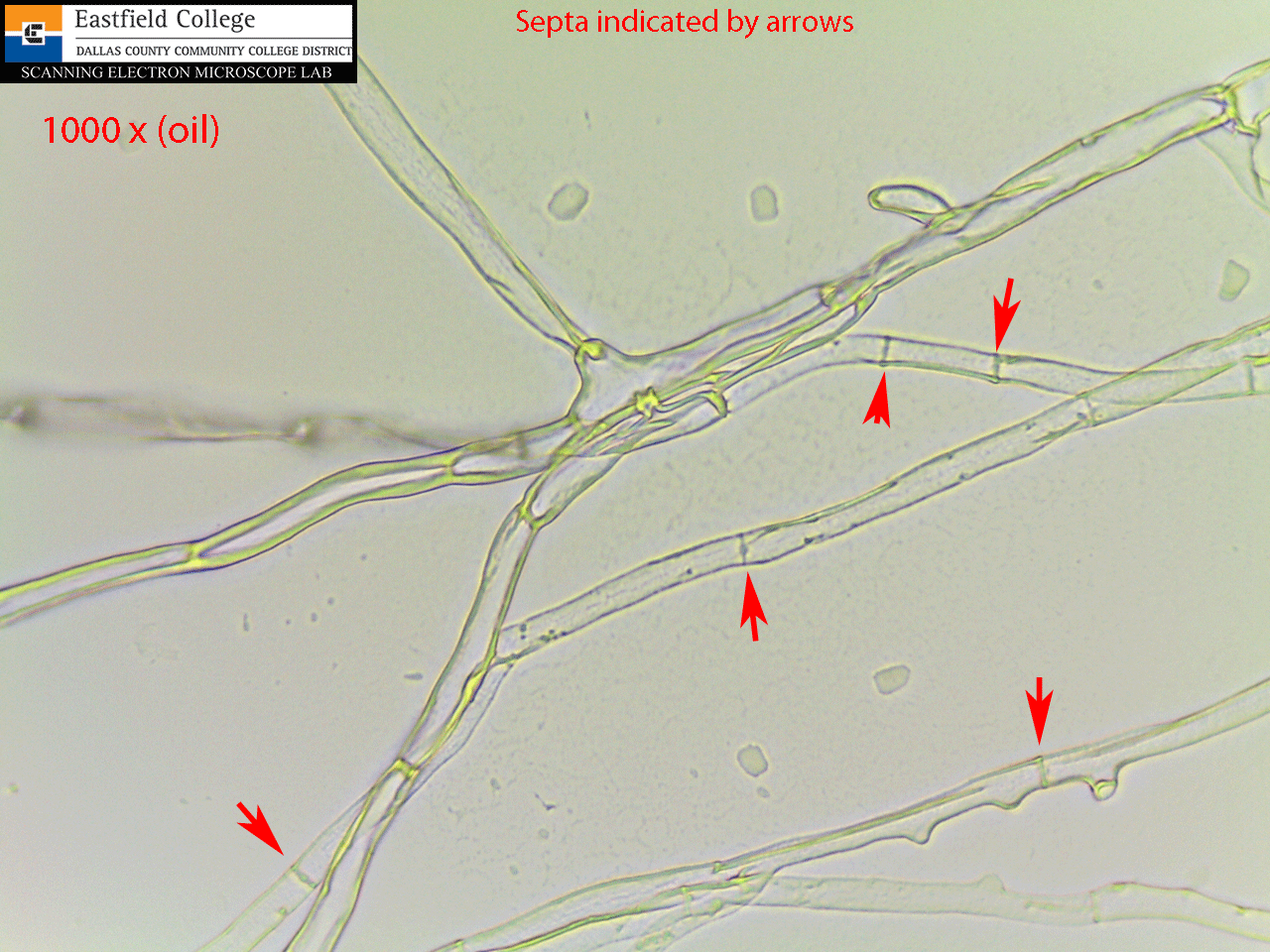
Morphology of the Fungi

Mold Hyphal Fragments - are Hyphae a Sign of Mold Contamination in  buildings?
Mold Hyphal Fragments - are Hyphae a Sign of Mold Contamination in buildings?

xanthocomically: An Introduction to Fungi The first thing to… – Roberts Lab

File:Fungal yeast cells, budding and hyphae in sputum specimen.jpg -  Wikimedia Commons
File:Fungal yeast cells, budding and hyphae in sputum specimen.jpg - Wikimedia Commons

Bacteria on the 'Fungal Highway': Pseudomonas putida moving along hyphae of  Cunninghamella elegans - YouTube
Bacteria on the 'Fungal Highway': Pseudomonas putida moving along hyphae of Cunninghamella elegans - YouTube

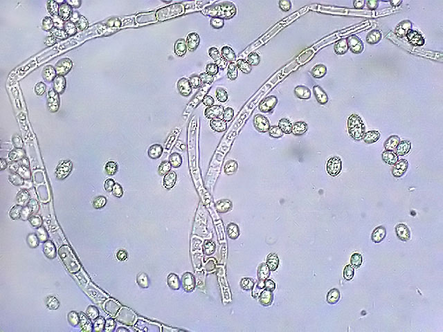
Scanning Electron Microscope Blog: April 2013

Life
Life

Hyphae hi-res stock photography and images - Alamy

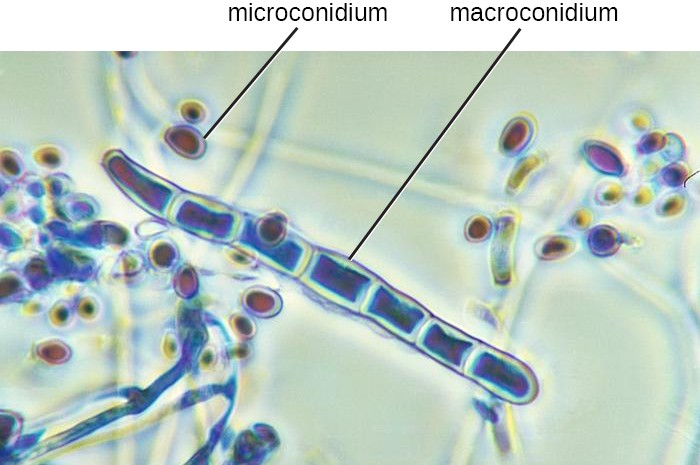
Pigmented fungal structures, fungal spores, hyphae and conidia (Olympus...  | Download Scientific Diagram
Pigmented fungal structures, fungal spores, hyphae and conidia (Olympus... | Download Scientific Diagram

Laboratory tests for fungal infections | DermNet

Fungi | Microbiology | | Course Hero

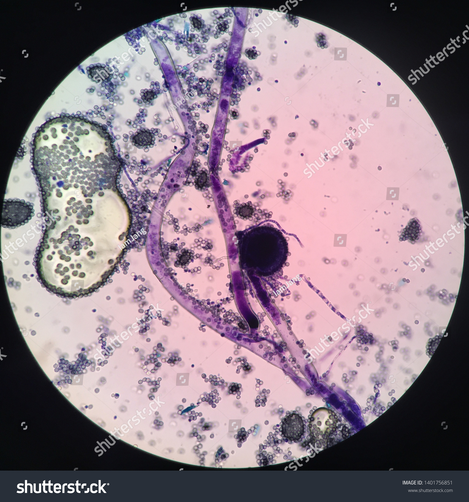
Aspergillus Spp Fungal Hyphae Under Microscope Stock Photo 1401756851 |  Shutterstock
Aspergillus Spp Fungal Hyphae Under Microscope Stock Photo 1401756851 | Shutterstock

Fungi Hyphae | Medical Laboratories

3,073 Fungal Hyphae Images, Stock Photos & Vectors | Shutterstock

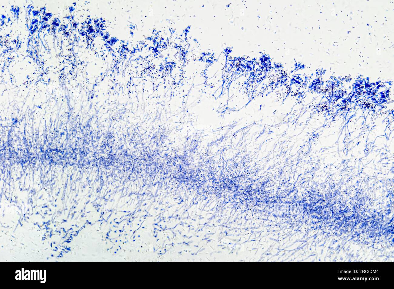
What is Mycelium? – Microscope Clarity