Home

Mâine gândac fond coccus electron microscope Biblie frigider Reporter

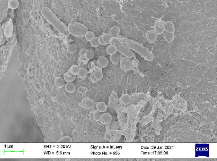
Scanning Electron Microscope (SEM) image of colony of coccus bacteria... |  Download Scientific Diagram
Scanning Electron Microscope (SEM) image of colony of coccus bacteria... | Download Scientific Diagram
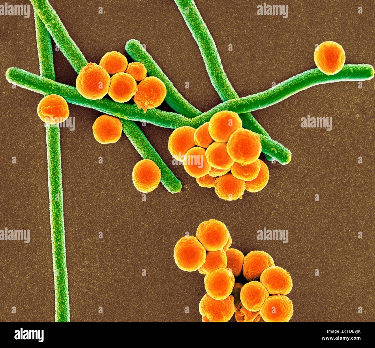
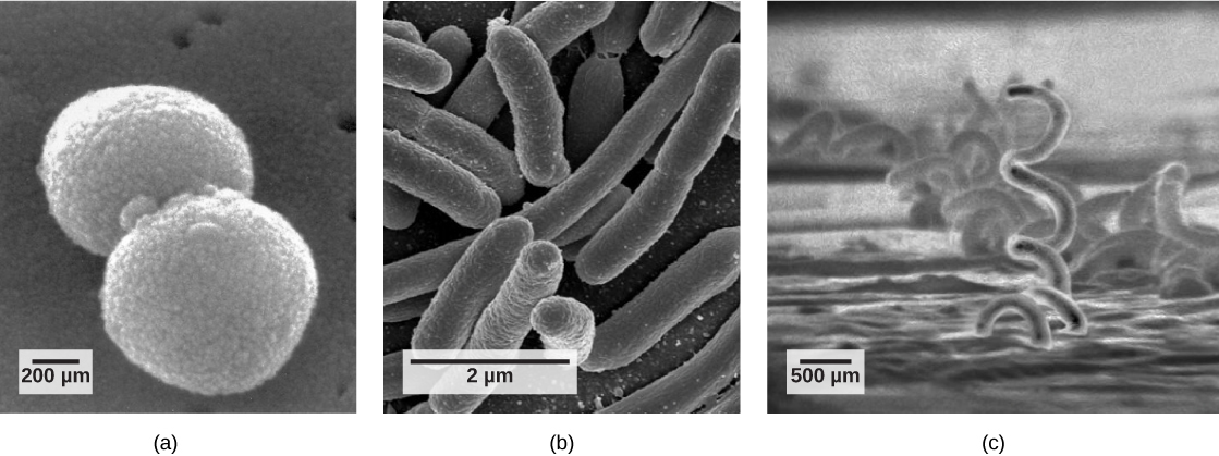
Coloured scanning electron micrograph (SEM) of rod-shaped (bacillus) and  round (coccus) bacteria Stock Photo - Alamy
Coloured scanning electron micrograph (SEM) of rod-shaped (bacillus) and round (coccus) bacteria Stock Photo - Alamy

PDF] Ultrastructure of the adhesion of bacteria to the epithelial cell  membrane of three-day postnatal rat tongue mucosa: a transmission and  high-resolution scanning electron microscopic study. | Semantic Scholar
PDF] Ultrastructure of the adhesion of bacteria to the epithelial cell membrane of three-day postnatal rat tongue mucosa: a transmission and high-resolution scanning electron microscopic study. | Semantic Scholar

Transmission electron microscope study of bacterial morphotypes on the  anterior dorsal surface of human tongues - Arora - 2000 - The Anatomical  Record - Wiley Online Library
Transmission electron microscope study of bacterial morphotypes on the anterior dorsal surface of human tongues - Arora - 2000 - The Anatomical Record - Wiley Online Library

Scanning Electron Micrograph (SEM) depicting large numbers of  Staphylococcus aureus bacteria, which were found on the luminal surface of  an indwelling catheter. A red blood cell (RBD), also known as an  erythrocyte,
Scanning Electron Micrograph (SEM) depicting large numbers of Staphylococcus aureus bacteria, which were found on the luminal surface of an indwelling catheter. A red blood cell (RBD), also known as an erythrocyte,

Free picture: scanning, electron micrograph, gram, positive, micrococcus  luteus, bacteria
Free picture: scanning, electron micrograph, gram, positive, micrococcus luteus, bacteria

Ultra-thin section of an uncultured magnetotactic coccus from microcosm...  | Download Scientific Diagram
Ultra-thin section of an uncultured magnetotactic coccus from microcosm... | Download Scientific Diagram
The Electron Microscopy Center of GTIIT - GUANGDONG TECHNION-ISRAEL  INSTITUTE OF TECHNOLOGY
The Electron Microscopy Center of GTIIT - GUANGDONG TECHNION-ISRAEL INSTITUTE OF TECHNOLOGY

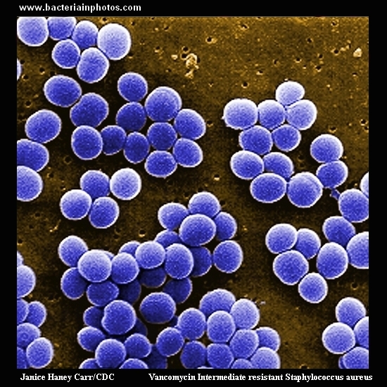
Scanning electron micrograph (SEM) of Staphylococcus aureus bacteria taken  from a vancomycin intermediate resistant culture (VISA)
Scanning electron micrograph (SEM) of Staphylococcus aureus bacteria taken from a vancomycin intermediate resistant culture (VISA)

Scanning electron microscope | iqb.rutgers.edu

Solved In the figure, identify which of the shapes are | Chegg.com

Scanning electron microscope image of co-isolated filamentous or... |  Download Scientific Diagram
Scanning electron microscope image of co-isolated filamentous or... | Download Scientific Diagram

Enterococcus faecalis, coccus prokaryote, SEM - Stock Image - C037/0113 -  Science Photo Library
Enterococcus faecalis, coccus prokaryote, SEM - Stock Image - C037/0113 - Science Photo Library

Rod-shaped (bacillus) and round (coccus) bacteria | Scanning electron  microscope, Electron microscope, Insect pollinators
Rod-shaped (bacillus) and round (coccus) bacteria | Scanning electron microscope, Electron microscope, Insect pollinators

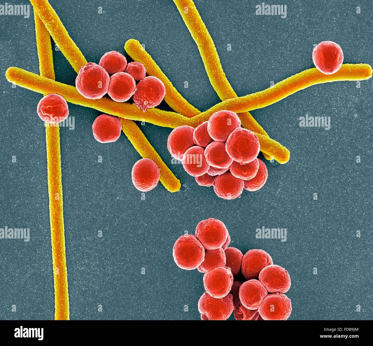
Staphylococcus aureus. Coloured scanning electron micrograph (SEM) of  Staphylococcus aureus bacteria. These Gram-positive bacteria cause skin  infections and often grow in these grape-like clusters of small spheres ( cocci). S. aureus is extremely
Staphylococcus aureus. Coloured scanning electron micrograph (SEM) of Staphylococcus aureus bacteria. These Gram-positive bacteria cause skin infections and often grow in these grape-like clusters of small spheres ( cocci). S. aureus is extremely

Biology, Biological Diversity, Prokaryotes: Bacteria and Archaea, Structure  of Prokaryotes | GoOpen CT
Biology, Biological Diversity, Prokaryotes: Bacteria and Archaea, Structure of Prokaryotes | GoOpen CT

Biology - Bacteria - Cocci seen through the scanning electron microscope,  Stock Photo, Picture And Rights Managed Image. Pic. DAE-10397875 |  agefotostock
Biology - Bacteria - Cocci seen through the scanning electron microscope, Stock Photo, Picture And Rights Managed Image. Pic. DAE-10397875 | agefotostock
NIAH:NIAH Pathogenic Organisms Observed by Electron Microscope:Coccidium

A) Light and (B–E) transmission electron microscopy micrographs of... |  Download Scientific Diagram
A) Light and (B–E) transmission electron microscopy micrographs of... | Download Scientific Diagram

History
History

3-D imaging could answer fundamental questions about valley fever | News |  bakersfield.com
3-D imaging could answer fundamental questions about valley fever | News | bakersfield.com

Coloured scanning electron micrograph (SEM) of rod-shaped (bacillus) and  round (coccus) bacteria Stock Photo - Alamy
Coloured scanning electron micrograph (SEM) of rod-shaped (bacillus) and round (coccus) bacteria Stock Photo - Alamy

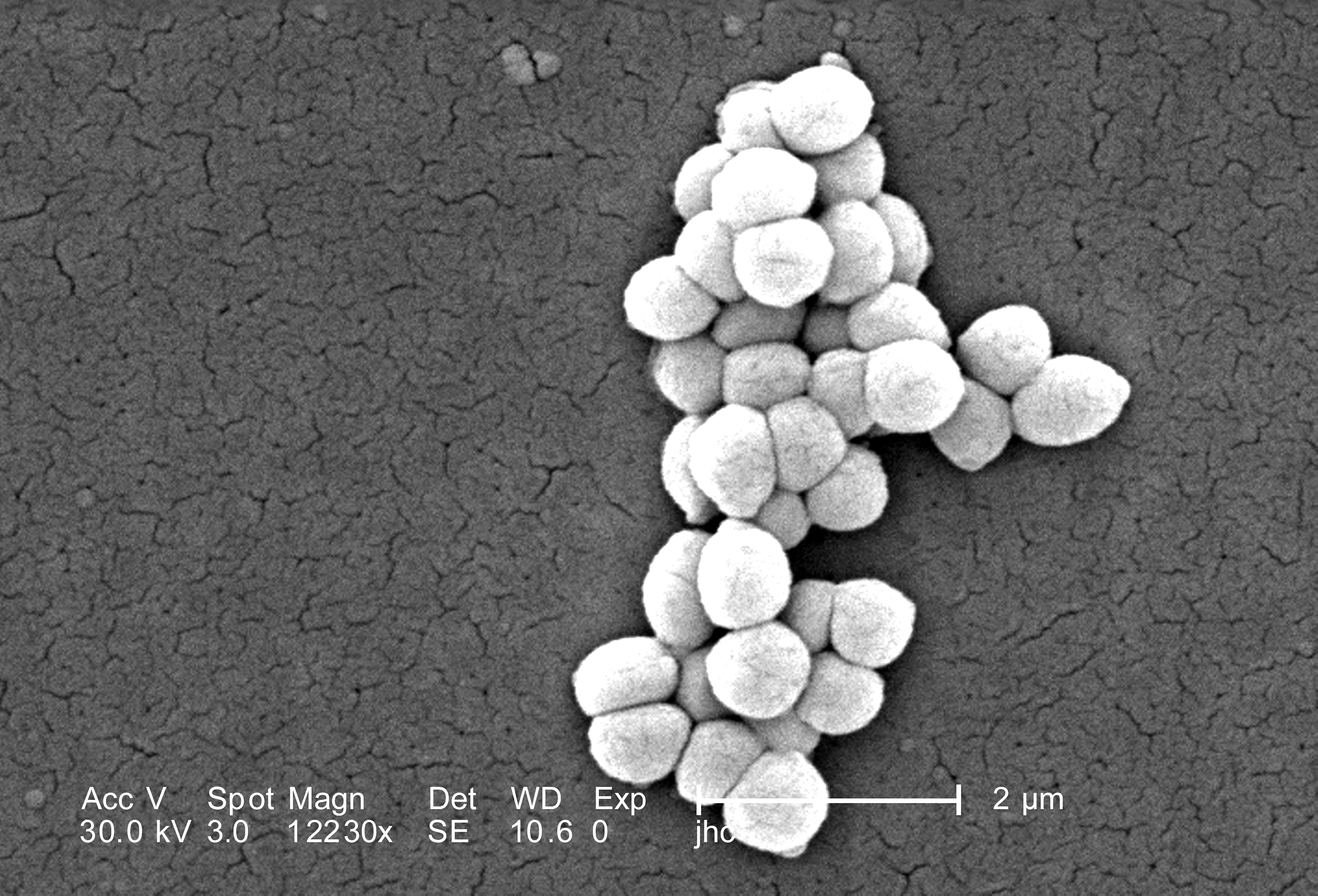
coccus stock photos - OFFSET

Solved A) Transmission Electron Microscopy Samples prepared | Chegg.com

IJMS | Free Full-Text | A Novel Ruthenium Based Coordination Compound  Against Pathogenic Bacteria | HTML
IJMS | Free Full-Text | A Novel Ruthenium Based Coordination Compound Against Pathogenic Bacteria | HTML